Found a Lump on Your Dog? A Calm 48-Hour Action Plan
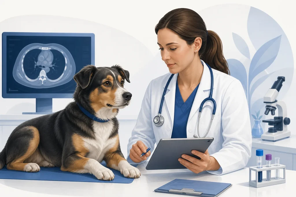
Your hand pauses during a belly rub: there is a bump that you do not remember feeling before. The mind naturally jumps to cancer. Take a breath. Many canine masses are benign, and some serious tumors can look ordinary. That is exactly why appearance, firmness, or mobility cannot settle the question. A prompt veterinary examination and, when appropriate, microscopic sampling are more useful than an anxious night of image searches.
First 10 minutes: look without provoking it
Note the precise location and whether the lump is on the skin, beneath it, or associated with a nipple, toe, mouth, or another structure. Look for redness, heat, hair loss, discharge, bruising, an open surface, or active bleeding. Avoid squeezing, puncturing, massaging, or applying creams. Some tumors—including mast cell tumors—can become swollen or irritated after manipulation, and any damaged lump can become infected or bleed.
Take one clear photo in good light with a ruler beside, not pressed into, the mass. A second photo from farther away can show the body location. Write the date. If your dog is comfortable, measure the longest visible dimension and the dimension across it. These observations help the veterinarian understand change; they do not identify the mass.
Next: check the whole dog
Ask whether anything else has changed: appetite, drinking, urination, bowel movements, sleep, energy, breathing, weight, gait, play, grooming, or social behavior. Check the gums only if your dog safely allows it. Pale or white gums, sudden weakness, collapse, a rapidly enlarging abdomen, breathing difficulty, uncontrolled bleeding, repeated vomiting, or severe distress warrants emergency veterinary care now—not a 48-hour watch.
A rapidly swelling face or muzzle, hives, or breathing changes may also represent an allergic emergency. A painful eye-area mass, inability to urinate or defecate, and a growth that obstructs eating deserve urgent advice. When uncertain, call a veterinary clinic and describe the signs rather than waiting for an online reply.
Within 24 hours: make the appointment
Contact your primary veterinarian about a new or changing mass. Tell the clinic if it is growing quickly, ulcerated, bleeding, painful, in the mouth, near an eye, affecting movement, or accompanied by general illness; that information helps triage the visit. Do not delay solely because the lump is small, soft, or movable. Cornell notes that benign and malignant skin masses can resemble one another and cannot be identified with complete certainty by sight and touch.
Prepare a complete list of medications, preventives, foods, treats, and supplements. Include photos of labels, amounts, and the time of the last dose. Also note previous tumors and pathology results. If your dog has had a similar-looking lipoma, remember that a new mass is a new question; resemblance does not establish identity.
At the visit: ask about sampling
The veterinarian may recommend a fine-needle aspirate, which collects cells for cytology and is often minimally invasive. Some samples provide a clear answer; others are nondiagnostic because the lesion sheds few cells or the sample is not representative. A biopsy obtains tissue and may provide the architecture, tumor grade, or other features needed for planning. Ask what the proposed test can answer, who will interpret it, and what the next step would be if it is inconclusive.
Before agreeing to simply remove a suspicious mass, ask whether biopsy or surgical planning is needed first. The ideal incision and margin can depend on tumor type and location. A planned definitive operation may preserve better options than an unplanned narrow removal.
While waiting: monitor consistently
Photograph no more often than needed to capture meaningful change, using the same angle and scale. Prevent licking if your veterinarian recommends a safe collar. Do not start leftover antibiotics, steroids, pain medicines, herbal preparations, or human products. These may be unsafe, interfere with tests, or mask changes. Call sooner if the mass changes rapidly or your dog develops systemic signs.
What not to conclude
- Soft does not automatically mean benign.
- Fast growth does not prove cancer, but it deserves prompt assessment.
- No pain does not make a mass harmless.
- A normal blood test does not identify most skin masses.
- A nondiagnostic aspirate is not a negative cancer test.
The goal of the first 48 hours is not to name the lump yourself. It is to preserve useful information, recognize emergencies, and move efficiently toward a veterinary diagnosis.
Medical disclaimer: This article is general education, not a diagnosis or an individual triage decision. A licensed veterinarian must examine and, when indicated, sample a mass. Seek emergency care for collapse, difficulty breathing, uncontrolled bleeding, pale gums, severe pain, or rapid deterioration.
Sources and further reading
- Cornell: Skin Lumps and Bumps
- AAHA: Tumor Diagnostics & Staging
- FDA: My Dog Has Cancer—What Do I Need to Know?
