Canine Cutaneous Squamous Cell Carcinoma: A Persistent Crust or Sore Needs Sampling
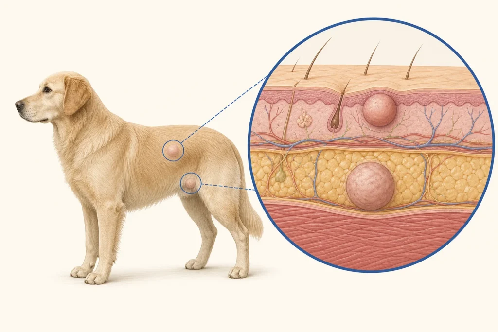
Overview
Cutaneous squamous cell carcinoma is a malignant tumor of epidermal cells. It may begin as a thickened, crusted plaque or a wound-like lesion that fails to heal, particularly on sparsely haired or chronically sun-exposed skin; infection and inflammatory disease can look similar.
Common signs
A persistent crust, raised plaque, firm nodule, ulcer, bleeding surface, discharge, odor, licking or local pain. Change despite routine wound care is an important clue, not proof of cancer.
Common locations
Sparsely haired skin of the trunk, abdomen, limbs, nose or other sun-exposed areas. Digital and oral squamous cell carcinomas behave differently and are covered separately.
Diagnosis discussion
Cytology can help exclude some mimics, but biopsy and histopathology usually establish the diagnosis and biologic features. The clinician may assess regional lymph nodes and select imaging when lesion size, location or pathology makes spread a meaningful concern.
Urgency and when to contact a veterinarian
Arrange a prompt appointment for any sore or crust that persists, enlarges, bleeds or repeatedly reopens. Seek urgent care for uncontrolled bleeding, severe pain, marked infection-like illness or rapid deterioration.
Treatment discussion
Complete planned surgical removal is often the main local-control discussion. Radiation or other local/systemic options may be considered when margins, location, recurrence or spread make surgery alone insufficient.
Home monitoring
Photograph beside a ruler, prevent licking as directed, and record bleeding, odor, pain and surface change. After treatment, inspect both the scar and regional nodes on the clinic's schedule.
Questions to ask your veterinarian
- Does this lesion need an incisional biopsy before definitive surgery?
- What margins and reconstruction should be planned for the first operation?
- Should the regional lymph node be sampled?
- Which pathology findings change follow-up or additional therapy?