Canine Digital Squamous Cell Carcinoma: When a Toe Lesion Is More Than a Nail Problem
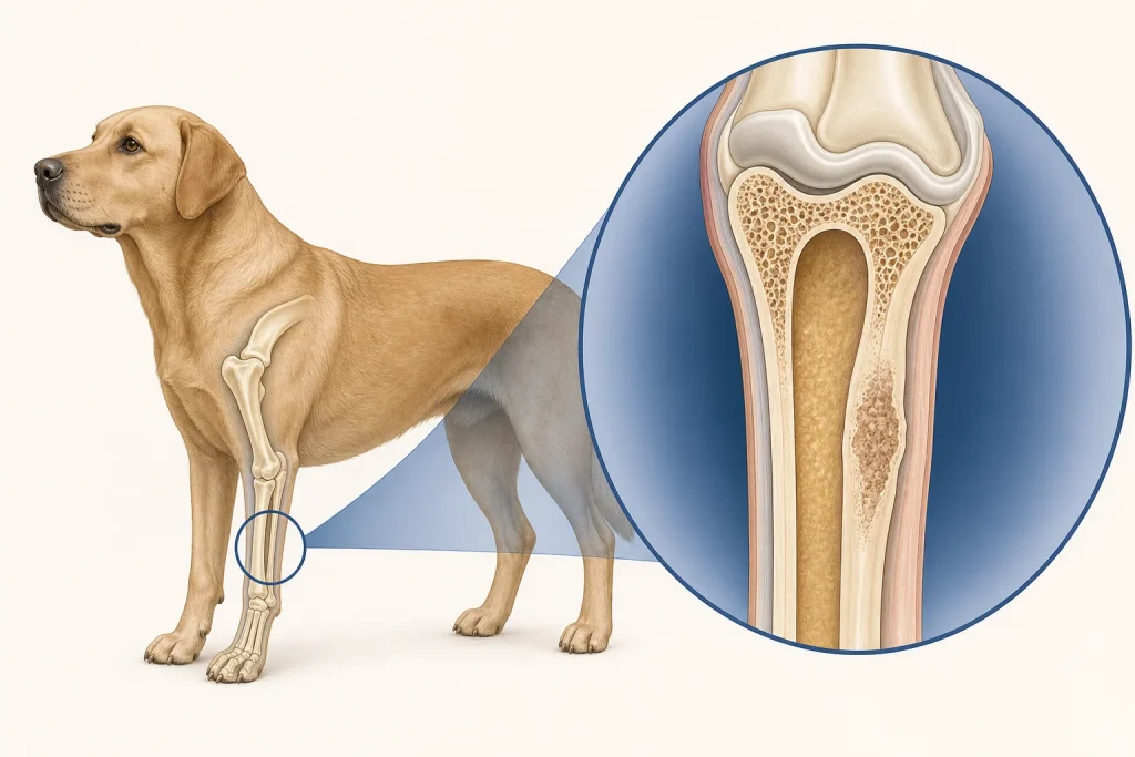
Overview
Digital squamous cell carcinoma can arise around the nail bed or toe and may first resemble a torn nail, infection or foreign body. Persistence, progressive swelling, nail loss or recurrent bleeding warrants a structured workup.
Common signs
Toe swelling, lameness, licking, a loose or missing nail, bloody discharge, ulceration, odor or pain when the foot is handled. Antibiotic response does not by itself prove or exclude cancer.
Common locations
Nail bed, skin of a toe, or adjacent digit tissues; nearby bone can become involved. More than one digit may require separate assessment rather than assumption.
Diagnosis discussion
Foot radiographs evaluate bone involvement, while cytology or biopsy provides tissue diagnosis. The veterinarian may sample the draining lymph node and recommend chest imaging or other staging based on histology and extent.
Urgency and when to contact a veterinarian
Book promptly for a toe or nail-bed problem that recurs or does not heal. Urgent care is appropriate for uncontrolled bleeding, severe pain, inability to bear weight, fever or rapidly spreading swelling.
Treatment discussion
Removal of the affected digit is a common local-control discussion when disease is confined and surgery is feasible. Pathology, margins, lymph-node findings and imaging guide whether additional treatment or surveillance is appropriate.
Home monitoring
Keep the foot clean and protected exactly as directed, restrict activity after surgery, and record gait, licking, discharge and bandage condition. Check other digits without squeezing lesions.
Questions to ask your veterinarian
- Should radiographs be taken before choosing the biopsy or surgery plan?
- Is digit amputation likely to provide adequate local control?
- Should the regional lymph node be sampled even if normal-sized?
- What bandage or incision changes require same-day review?