Canine Sebaceous Gland Tumors: Benign-Looking Growths Still Deserve Identification
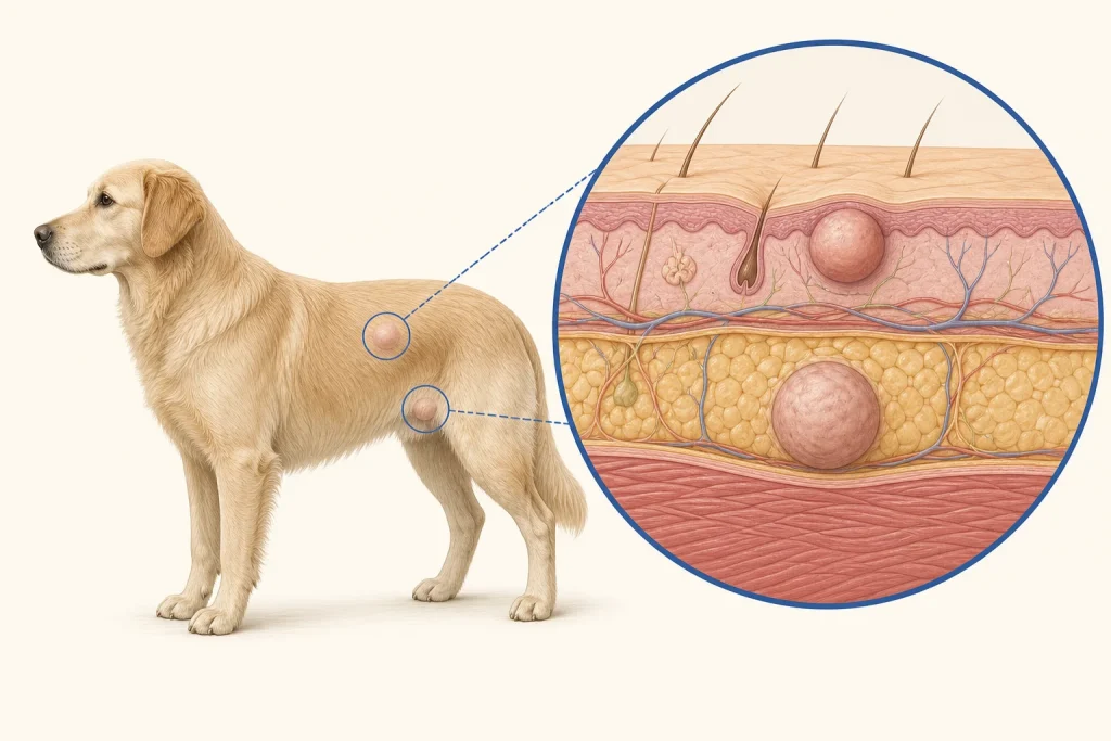
Overview
Sebaceous gland tumors arise from oil-producing structures in the skin. Many are benign adenomas or epitheliomas, but appearance overlaps with other follicular, glandular and malignant lesions, so a visual label is not enough.
Common signs
A raised, lobulated or wart-like bump that may be pink, tan or pigmented; crusting, minor bleeding and irritation can occur when a collar, grooming tool or licking traumatizes it.
Common locations
Head, eyelid-adjacent facial skin, trunk and limbs are common sites. A lesion near the eye or other delicate structure needs site-specific planning.
Diagnosis discussion
Fine-needle aspiration may narrow the possibilities, but biopsy or excision with histopathology gives the most reliable classification. Faster growth, fixation, ulceration or recurrence increases the value of tissue diagnosis.
Urgency and when to contact a veterinarian
Most small stable lesions can be seen at a prompt routine appointment. Request earlier care for rapid growth, repeated bleeding, infection-like change, pain or interference with vision, eating or movement.
Treatment discussion
Observation may be reasonable after a veterinarian confirms a low-risk diagnosis and sets a recheck plan. Surgical removal is considered for uncertainty, irritation, functional interference or growth; pathology should name the lesion.
Home monitoring
Use a body map and consistent photographs. Record diameter, surface crust, bleeding, licking and whether the lesion catches on grooming equipment or a collar.
Questions to ask your veterinarian
- Can cytology reasonably identify this lesion, or is biopsy preferable?
- Would removal now be simpler than after further growth?
- Will the entire sample be submitted for histopathology?
- What specific change should trigger an earlier recheck?