Canine Cutaneous Melanocytic Tumors: Pigment Does Not Predict Behavior
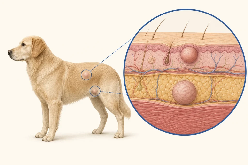
Overview
Melanocytic tumors arise from pigment-producing cells and range from benign melanocytomas to malignant melanoma. Color alone is unreliable: some benign lesions are dark, some malignant lesions have little pigment, and behavior differs strongly by anatomic site.
Common signs
A dark, gray, pink or mixed-color nodule; enlargement, ulceration, bleeding or fixation can occur. Many are found during grooming before they affect comfort.
Common locations
Haired skin, lips, nail bed and oral cavity. This page focuses on haired-skin lesions; oral and digital tumors require their own risk discussion.
Diagnosis discussion
Fine-needle aspiration may suggest a melanocytic tumor, but histopathology is needed for classification, margins and features that inform risk. Regional-node assessment and staging are selected according to site and pathology.
Urgency and when to contact a veterinarian
Arrange a prompt appointment for any new pigmented or changing mass. Earlier care is warranted for rapid growth, ulceration, bleeding, pain, a nail-bed location or an oral lesion.
Treatment discussion
Complete surgery is often the first local-control option for a resectable skin tumor. Additional local or systemic therapy depends on anatomic site, histologic findings, margins, recurrence and evidence of spread.
Home monitoring
Photograph with a ruler and record size, color, surface change, bleeding and nearby node enlargement. Do not interpret fading or darkening as proof of response.
Questions to ask your veterinarian
- Does cytology support a melanocytic lesion, and is biopsy or excision next?
- How does this exact anatomic site affect expected behavior?
- What do margins and pathology features mean for staging?
- Should the regional lymph node be sampled?