Canine Infundibular Keratinizing Acanthoma: A Pore-Like Mass Can Mimic Other Tumors
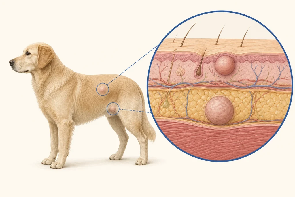
Overview
Infundibular keratinizing acanthoma is a usually benign follicular tumor that forms a keratin-filled crater or pore. It may resemble a cyst, wound or other follicular tumor and can become inflamed if it ruptures or is manipulated.
Common signs
A firm, raised nodule with a central opening or plug of keratin, sometimes with crust, discharge, redness or licking. Multiple lesions can occur in predisposed dogs.
Common locations
Trunk, neck and limbs are common sites in the skin. Depth and multiplicity influence removal and monitoring plans.
Diagnosis discussion
Cytology may recover keratin but cannot always distinguish a cyst from a follicular neoplasm. Histopathology after biopsy or excision confirms the architecture and excludes a more concerning look-alike.
Urgency and when to contact a veterinarian
A small quiet lesion can be assessed at a prompt appointment. Earlier care is needed for painful swelling, repeated rupture, odor, spreading redness, rapid enlargement or persistent bleeding.
Treatment discussion
Complete surgical excision is generally curative for a solitary confirmed lesion. Multiple lesions require an individualized plan balancing symptoms, diagnostic certainty and procedure burden.
Home monitoring
Do not squeeze or lance the opening. Photograph and measure it, record rupture or discharge, and monitor excision sites for a new central plug or regrowth.
Questions to ask your veterinarian
- Is the sample diagnostic, or could this still be another follicular lesion?
- Would intact removal improve diagnosis and comfort?
- If there are multiple lesions, which should be sampled first?
- What post-removal change suggests recurrence?