Canine Ceruminous Gland Tumors: Chronic Ear Discharge May Hide a Mass
Overview
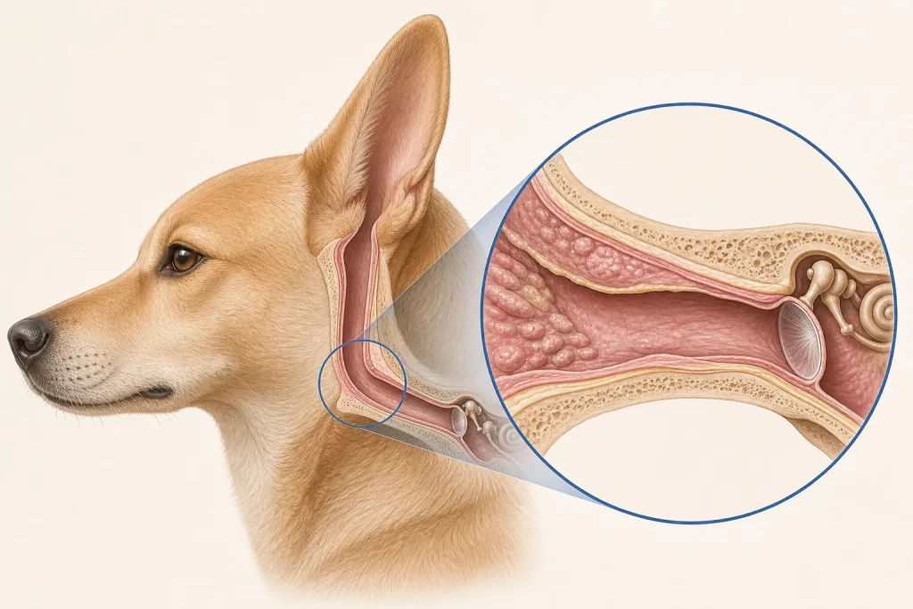
Ceruminous gland tumors arise from wax-producing glands of the external ear canal. Benign and malignant forms can both narrow the canal and promote chronic inflammation, so repeated treatment of discharge without visualizing the canal can delay diagnosis.
Common signs
One-sided chronic discharge, odor, head shaking, scratching, pain, bleeding, reduced hearing or a visible canal mass. Secondary infection may coexist.
Common locations
External ear canal, from the opening toward the eardrum. Extent toward the middle ear and surrounding tissues is important for surgical planning.
Diagnosis discussion
A careful ear examination under appropriate sedation or anesthesia may reveal the lesion. Cytology evaluates infection and some cells, while biopsy establishes tumor type. CT or other imaging may be recommended to map deeper disease.
Urgency and when to contact a veterinarian
Arrange prompt assessment for persistent one-sided ear disease, bleeding or a visible mass. Severe pain, loss of balance, facial weakness, inability to eat or rapidly worsening neurologic signs needs urgent care.
Treatment discussion
Small accessible benign tumors may be locally removed; more extensive or malignant tumors can require wider ear-canal surgery and staging. Infection and pain should be treated alongside the tumor plan.
Home monitoring
Do not insert swabs or cleaners unless directed. Track discharge, odor, head tilt, balance, facial symmetry, appetite and pain, and keep postoperative wound checks.
Questions to ask your veterinarian
- Can the entire canal and eardrum be examined safely?
- Would CT change the biopsy or surgical plan?
- What does pathology say about invasion and margins?
- Which balance or facial-nerve signs require urgent contact?