Canine Fibrosarcoma: Plan the First Surgery Around Microscopic Extension
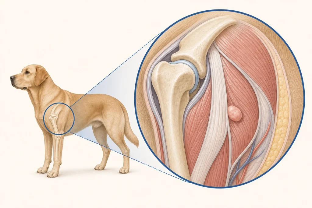
Overview
Fibrosarcoma is a malignant connective-tissue tumor within the broader soft-tissue sarcoma family. It may feel discrete while microscopic cells extend beyond what fingers or imaging can define, making preoperative diagnosis and margin planning important.
Common signs
A firm or deep mass that gradually enlarges, becomes fixed, causes pressure or pain, or interferes with movement. The overlying skin can remain normal until the mass is large.
Common locations
Skin, subcutaneous tissue, muscle and connective tissue of the trunk, limbs, head or oral cavity. Site influences resectability and behavior.
Diagnosis discussion
Fine-needle aspiration may be inconclusive for mesenchymal tumors. Core or incisional biopsy can establish the tumor family and guide definitive surgery. Imaging helps map deep or fixed masses; histopathology reports grade and margins.
Urgency and when to contact a veterinarian
Arrange a prompt appointment for every persistent deep or growing mass. Urgent evaluation is appropriate for rapid functional decline, severe pain, ulceration, uncontrolled bleeding or breathing difficulty.
Treatment discussion
Wide planned surgery is a common local-control approach. Radiation may be discussed for difficult sites or residual microscopic disease; systemic therapy is selected by grade, stage and individual risk.
Home monitoring
Measure in two directions, photograph, and record firmness, mobility, gait or posture effects and pain. After treatment, monitor the scar and nearby tissue rather than only the original center.
Questions to ask your veterinarian
- Should a core or incisional biopsy occur before definitive surgery?
- What imaging is needed to map deep extension?
- What margins are realistic at this location?
- How do grade and margins shape radiation, staging and rechecks?