Canine Eyelid Tumors: Protecting the Eye While Naming the Mass
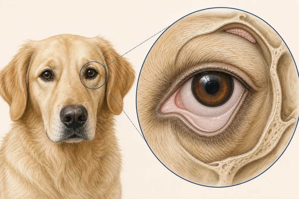
Overview
Eyelid tumors are common in dogs and many are benign, but even a small mass can rub the cornea or distort normal blinking. Appearance cannot reliably separate every benign lesion from a malignant or locally invasive tumor.
Common signs
A new lid-edge bump, change in pigment, bleeding, crusting, rubbing, squinting, tearing, discharge, or incomplete eyelid closure may be seen. Eye pain can be subtle and should not be judged only by whether the dog is still eating.
Common locations
Masses may arise from glands along the lid margin, skin, deeper lid tissue, or the third eyelid. Their position relative to the tear ducts, cornea, and lid margin influences surgical reconstruction.
Diagnosis discussion
A complete eye examination checks the cornea and other ocular structures. Cytology can help in selected lesions, while excision or biopsy with histopathology names the tumor and evaluates margins. Planning before removal helps preserve eyelid function.
Urgency and when to contact a veterinarian
Squinting, a blue or cloudy cornea, sudden vision concern, marked swelling, trauma, or uncontrolled bleeding needs same-day veterinary advice. A quiet, small eyelid mass still deserves a prompt planned examination.
Treatment discussion
Surgery is commonly discussed when a lesion irritates the eye, grows, or is diagnostically uncertain. Technique depends on size and location; referral to ophthalmology or surgery may protect eyelid closure and the ocular surface. Additional therapy depends on pathology.
Home monitoring
Photograph the closed and open eye without touching the mass. Track rubbing, blink completeness, tearing, discharge, redness, mass growth, and prescribed eye-medication use. Prevent self-trauma only with veterinarian-approved protection.
Questions to ask your veterinarian
- Is the cornea already being irritated?
- Can the mass be removed while preserving lid function?
- Will the entire sample be submitted for histopathology and margin review?
- Should an ophthalmologist or reconstructive surgeon be involved before the first procedure?