Canine Subcutaneous Mast Cell Tumors: Location Below the Skin Matters
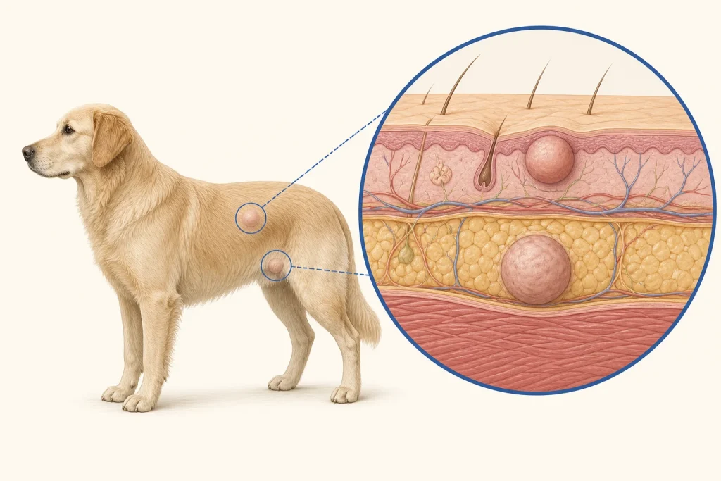
Overview
A mast cell tumor centered in the subcutaneous fat is biologically and pathologically assessed differently from one arising in the dermis. A soft movable feel does not distinguish it from lipoma or another subcutaneous tumor.
Common signs
A lump under apparently normal skin may enlarge slowly, fluctuate, become red or bruised, itch, or remain quiet. Vomiting, poor appetite, or dark stool can occur with mast-cell mediator effects but are not present in every dog.
Common locations
Subcutaneous mast cell tumors can occur on the trunk, limbs, head, or neck. Depth, fascial involvement, and proximity to joints or major structures affect surgical planning.
Diagnosis discussion
Fine-needle aspiration often identifies mast cells. Histopathology should specify that the tumor is subcutaneous and report features relevant to risk and margins. Regional lymph-node sampling and selective staging are based on pathology and clinical context.
Urgency and when to contact a veterinarian
Collapse, breathing difficulty, marked acute swelling, repeated vomiting, black stool, or uncontrolled bleeding needs urgent care. Do not squeeze or repeatedly manipulate a suspected mast cell tumor.
Treatment discussion
Planned surgery is common for local control. Radiation or systemic therapy may be discussed for incomplete control, recurrence, high-risk features, nodal disease, or inaccessible anatomy. Decisions should use the specific pathology, not a generic mast-cell label.
Home monitoring
Photograph and measure without squeezing. Record swelling episodes, skin color, licking, appetite, vomiting, stool color, energy, drug effects, incision healing, and new lumps. Each new mass deserves its own sampling decision.
Questions to ask your veterinarian
- Does pathology confirm a subcutaneous rather than dermal origin?
- Which histologic features inform recurrence or spread risk?
- Was the relevant regional lymph node assessed?
- Are the surgical margins adequate for this specific site and report?