Canine Esophageal Tumors: Regurgitation Requires Localization, Not Guesswork
Overview
Esophageal Tumors are rare growths arising in or compressing the tube that carries food from the throat to the stomach. Different cancers and noncancer disorders can narrow or disrupt the esophagus, and regurgitation must be distinguished from vomiting because the diagnostic paths differ.
Common signs
Owners may notice passive return of food or water, repeated swallowing, drooling, discomfort when swallowing, weight loss, reduced appetite, cough, fever, or breathing change if material is aspirated. None of these signs is specific to esophageal tumors, so a veterinary examination and appropriate testing are needed before naming the problem.
Common locations
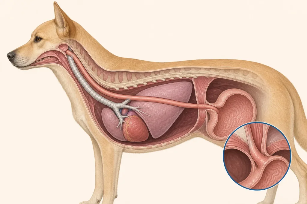
The esophagus travels through the neck and chest to the stomach; a lesion may be inside the wall, within the lumen, or compressing it from nearby tissue. Anatomy affects what can be sampled safely, whether a complete first surgery is realistic, and which nearby function needs protection.
Diagnosis discussion
History focused on regurgitation versus vomiting, chest imaging, contrast studies in selected cases, endoscopy, CT, and carefully planned biopsy may be used. The team also checks for aspiration pneumonia and nutritional compromise. The diagnostic plan should answer a decision: whether tissue type, local extent, stage, or patient health will change the next option. An imaging impression is not the same as a histologic diagnosis.
Urgency and when to contact a veterinarian
Arrange a prompt veterinary appointment for persistent or progressive signs. Seek emergency veterinary care for any of these signs: difficulty breathing, blue or gray gums, repeated inability to keep water down, marked weakness, collapse, or suspected aspiration pneumonia. Call ahead when possible so the receiving team can prepare; do not delay emergency transport to take photographs or complete a log.
Treatment discussion
Surgery, endoscopic procedures, radiation, systemic therapy, feeding-tube support, or palliative management may be considered depending on histology, length, location, spread, and aspiration risk. No food-texture change should replace diagnostic evaluation. The team should explain the goal of each option—diagnosis, local control, systemic control, symptom relief, or emergency stabilization—along with likely burdens, costs, travel, rechecks, and alternatives.
Home monitoring
Keep one dated home record of the timing and appearance of regurgitation, ability to keep water down, cough, respiratory rate at rest, fever if measured as instructed, meal texture, weight, hydration, and energy. Use the same measurement or observation method each time and share trends with the veterinary team. Home tracking supports planned rechecks; it cannot determine grade, margins, or metastasis.
Questions to ask your veterinarian
- Are these episodes regurgitation, vomiting, coughing, or a combination?
- Which imaging or endoscopic test best localizes the obstruction?
- How will biopsy risk and esophageal healing be managed?
- What nutrition plan limits aspiration while results are pending?