Canine Rectal Polyps and Adenomas: Benign Histology Still Can Affect Function
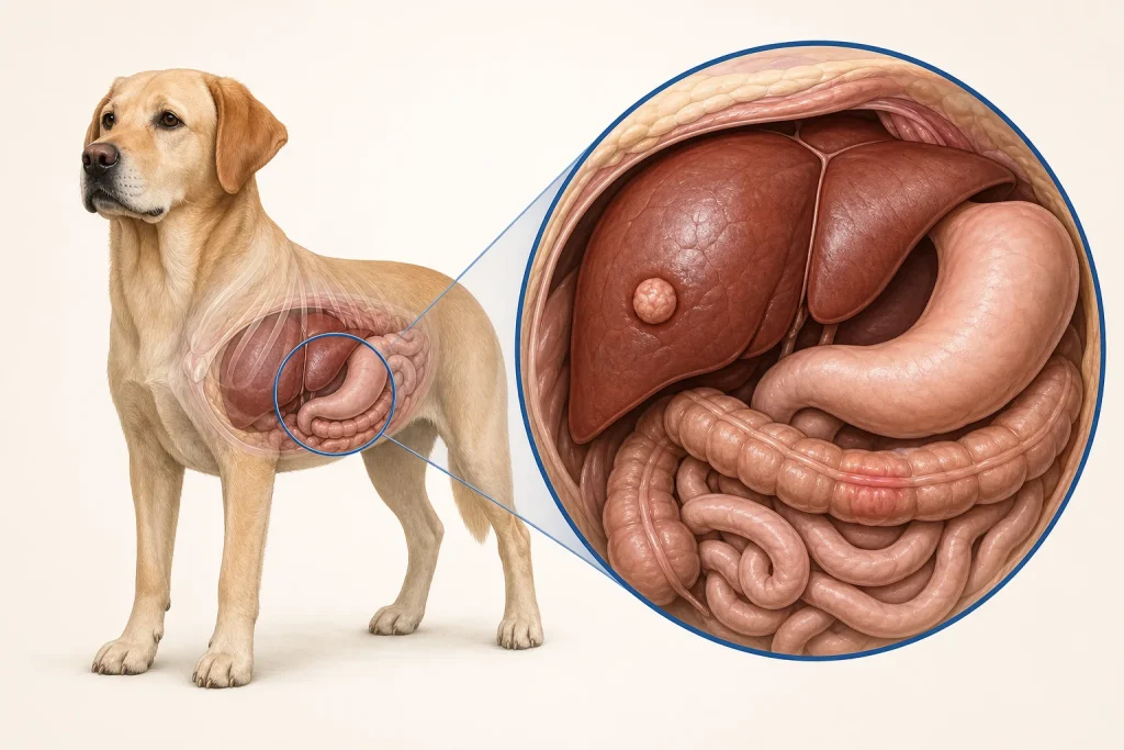
Overview
Rectal Polyps and Adenomas are mucosal growths that project into the rectum and may be benign adenomas, inflammatory polyps, or lesions with malignant change. Their surface appearance is not reliable enough to determine type, and even a benign lesion can bleed, recur, or obstruct defecation.
Common signs
Owners may notice fresh blood on stool, mucus, straining, frequent attempts to defecate, scooting, a protruding tissue mass, narrow stool, or discomfort when passing stool. None of these signs is specific to rectal polyps and adenomas, so a veterinary examination and appropriate testing are needed before naming the problem.
Common locations
Most are found within the rectal lumen, but the distance from the anus, number of lesions, attachment, and circumferential involvement shape treatment options. Anatomy affects what can be sampled safely, whether a complete first surgery is realistic, and which nearby function needs protection.
Diagnosis discussion
A rectal examination, endoscopic visualization, full-thickness or representative biopsy, and assessment for additional lesions may be recommended. Histopathology clarifies adenoma, inflammation, carcinoma, and margin status. The diagnostic plan should answer a decision: whether tissue type, local extent, stage, or patient health will change the next option. An imaging impression is not the same as a histologic diagnosis.
Urgency and when to contact a veterinarian
Arrange a prompt veterinary appointment for persistent or progressive signs. Seek urgent veterinary care for any of these signs: heavy bleeding, inability to pass stool, repeated vomiting with abdominal pain, collapse, marked weakness, or tissue that remains prolapsed and traumatized. Call ahead when possible so the receiving team can prepare; do not delay emergency transport to take photographs or complete a log.
Treatment discussion
Endoscopic, transanal, or other surgical removal may be considered depending on size and position. Follow-up examination matters because recurrence or additional polyps can occur, and malignant pathology changes staging and treatment planning. The team should explain the goal of each option—diagnosis, local control, systemic control, symptom relief, or emergency stabilization—along with likely burdens, costs, travel, rechecks, and alternatives.
Home monitoring
Keep one dated home record of blood or mucus, stool shape, time spent straining, frequency, prolapse-like tissue, pain, appetite, weight, and recurrence of the original pattern after removal. Use the same measurement or observation method each time and share trends with the veterinary team. Home tracking supports planned rechecks; it cannot determine grade, margins, or metastasis.
Questions to ask your veterinarian
- Was the sample deep and representative enough to exclude invasive carcinoma?
- Can the lesion be removed while preserving continence and rectal function?
- Were margins assessed, and what follow-up schedule is appropriate?
- What recurrence signs should prompt examination before the planned recheck?