Canine Parathyroid Tumors: High Calcium Can Be the First Clue
Overview
Parathyroid Tumors are growths of tiny hormone-producing glands near the thyroid that can release excess parathyroid hormone. Many are adenomas, but the urgent clinical issue is often hypercalcemia and its effect on kidneys, muscles, the gastrointestinal tract, and nervous system rather than the size of a neck nodule.
Common signs
Owners may notice increased thirst and urination, appetite change, vomiting, constipation, weakness, tremors, lethargy, urinary stones, or an unexpectedly high calcium result on bloodwork. None of these signs is specific to parathyroid tumors, so a veterinary examination and appropriate testing are needed before naming the problem.
Common locations
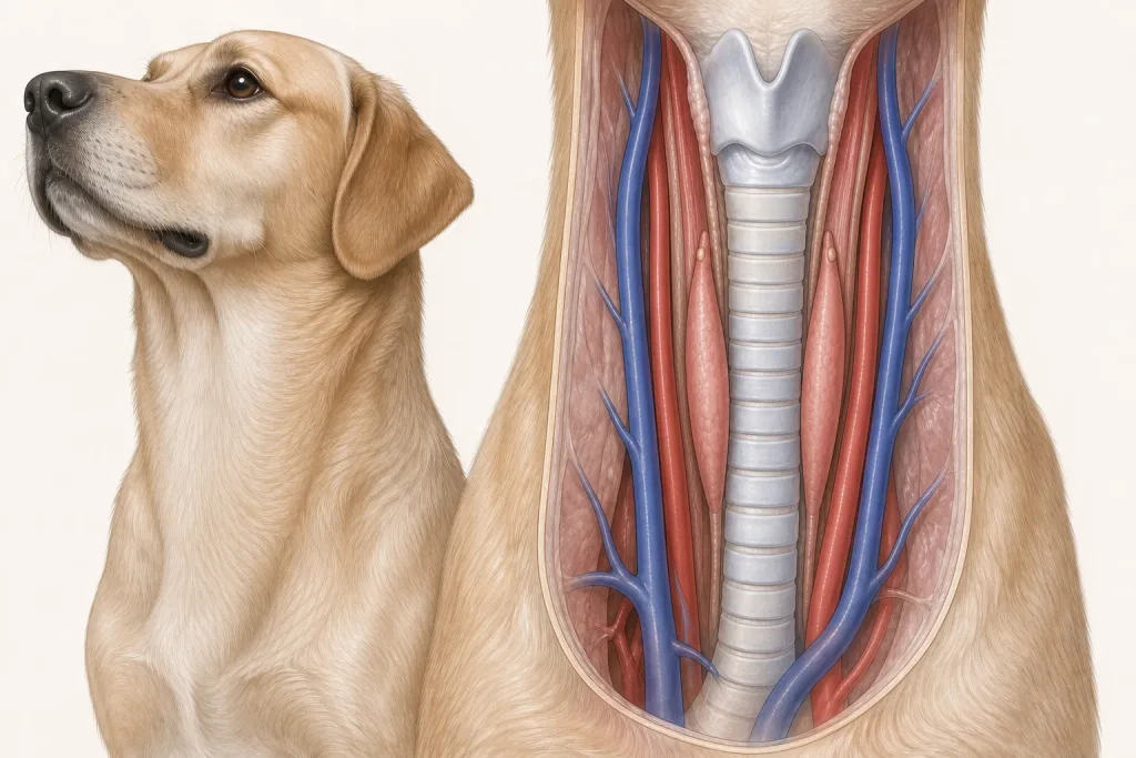
Parathyroid glands usually lie beside or within thyroid tissue in the neck, though ectopic tissue can occur; lesions may be too small to feel. Anatomy affects what can be sampled safely, whether a complete first surgery is realistic, and which nearby function needs protection.
Diagnosis discussion
Repeat total and ionized calcium, kidney and urine testing, parathyroid hormone interpretation, neck ultrasound, and evaluation for other causes of hypercalcemia may be needed. A high calcium value alone does not identify the cause. The diagnostic plan should answer a decision: whether tissue type, local extent, stage, or patient health will change the next option. An imaging impression is not the same as a histologic diagnosis.
Urgency and when to contact a veterinarian
Arrange a prompt veterinary appointment for persistent or progressive signs. Seek urgent veterinary care for any of these signs: severe weakness, repeated vomiting, inability to keep water down, collapse, seizures, marked tremors, or inability to urinate. Call ahead when possible so the receiving team can prepare; do not delay emergency transport to take photographs or complete a log.
Treatment discussion
Surgical removal, ultrasound-guided ablation in selected centers, or medical stabilization may be discussed. Calcium can fall after successful treatment, so planned postoperative monitoring and supplementation decisions belong to the veterinary team. The team should explain the goal of each option—diagnosis, local control, systemic control, symptom relief, or emergency stabilization—along with likely burdens, costs, travel, rechecks, and alternatives.
Home monitoring
Keep one dated home record of water intake, urine volume, appetite, vomiting, constipation, weakness, twitching, gait, and laboratory recheck dates rather than trying to assess a tiny neck lesion at home. Use the same measurement or observation method each time and share trends with the veterinary team. Home tracking supports planned rechecks; it cannot determine grade, margins, or metastasis.
Questions to ask your veterinarian
- Was ionized calcium confirmed and interpreted with parathyroid hormone?
- Which competing causes of hypercalcemia remain possible?
- Can imaging localize the abnormal gland well enough to plan treatment?
- How often will calcium be checked after treatment, and what signs suggest it is too low?