Canine Aortic Body Tumor: A Heart-Base Mass Can Affect Circulation or Breathing
Overview
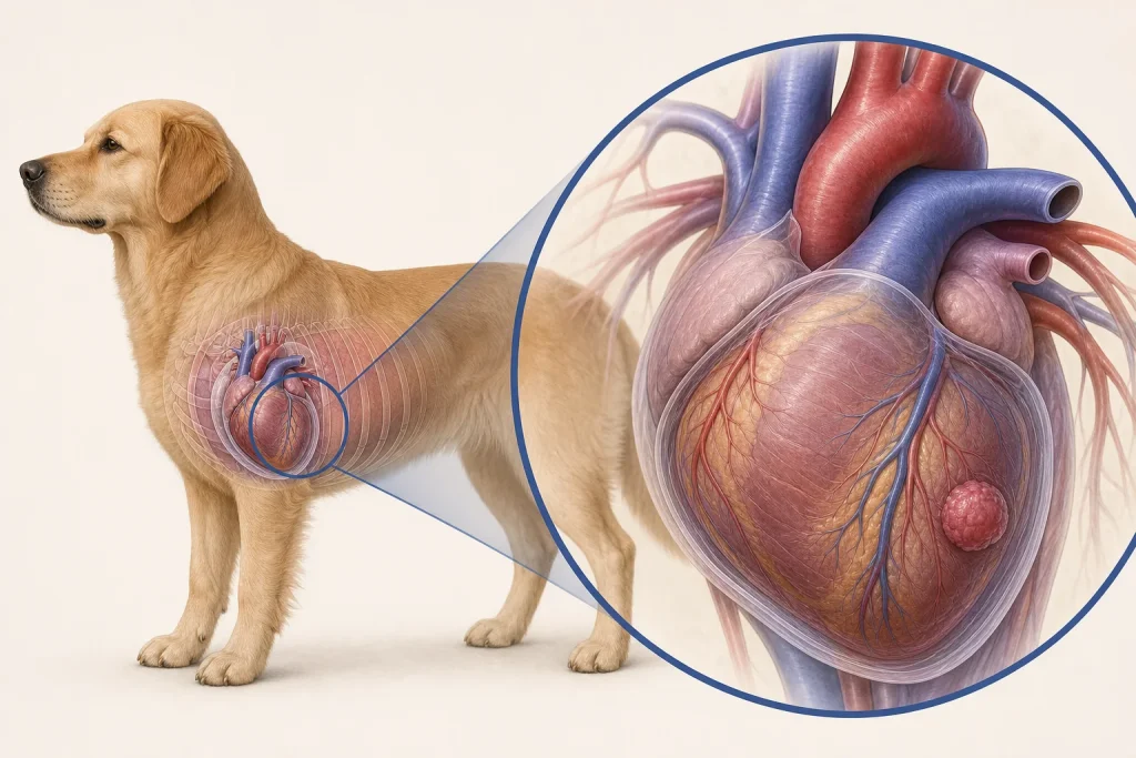
Aortic Body Tumor is a neuroendocrine tumor, also called a chemodectoma, arising from chemoreceptor tissue near the base of the heart. Some grow slowly, yet their position can compress vessels, airways, or the heart and can be associated with pericardial fluid; imaging cannot always provide definitive histology.
Common signs
Owners may notice exercise intolerance, cough, rapid or labored breathing, fainting, weakness, abdominal fluid, reduced appetite, or no signs when a heart-base mass is found incidentally. None of these signs is specific to aortic body tumor, so a veterinary examination and appropriate testing are needed before naming the problem.
Common locations
The mass typically develops near the aorta and pulmonary artery at the heart base inside the chest, where even noninvasive growth can affect critical structures. Anatomy affects what can be sampled safely, whether a complete first surgery is realistic, and which nearby function needs protection.
Diagnosis discussion
Echocardiography, chest radiographs or CT, blood pressure and cardiac evaluation, assessment for pericardial effusion, and staging may be recommended. Sampling can be high risk and is not always required before a carefully reasoned plan. The diagnostic plan should answer a decision: whether tissue type, local extent, stage, or patient health will change the next option. An imaging impression is not the same as a histologic diagnosis.
Urgency and when to contact a veterinarian
Arrange a prompt veterinary appointment for persistent or progressive signs. Seek emergency veterinary care for any of these signs: collapse, pale or blue gums, marked breathing effort, repeated fainting, a rapidly enlarging abdomen, or profound weakness. Call ahead when possible so the receiving team can prepare; do not delay emergency transport to take photographs or complete a log.
Treatment discussion
Observation with structured imaging, radiation, systemic therapy in selected cases, pericardial procedures, surgery in uncommon selected circumstances, or comfort-focused care may be discussed according to symptoms, growth, and anatomy. The team should explain the goal of each option—diagnosis, local control, systemic control, symptom relief, or emergency stabilization—along with likely burdens, costs, travel, rechecks, and alternatives.
Home monitoring
Keep one dated home record of sleeping respiratory rate, breathing effort, cough, fainting or weakness episodes, exercise tolerance, abdominal size, appetite, and the scheduled imaging comparison. Use the same measurement or observation method each time and share trends with the veterinary team. Home tracking supports planned rechecks; it cannot determine grade, margins, or metastasis.
Questions to ask your veterinarian
- Which imaging features support a heart-base origin rather than another mediastinal mass?
- Is pericardial fluid present, and what recurrence plan is needed?
- Would tissue sampling change management enough to justify its risk?
- Which home breathing or collapse signs require immediate transport?