Canine Multilobular Tumor of Bone: A Slow Skull Mass Can Still Become Invasive
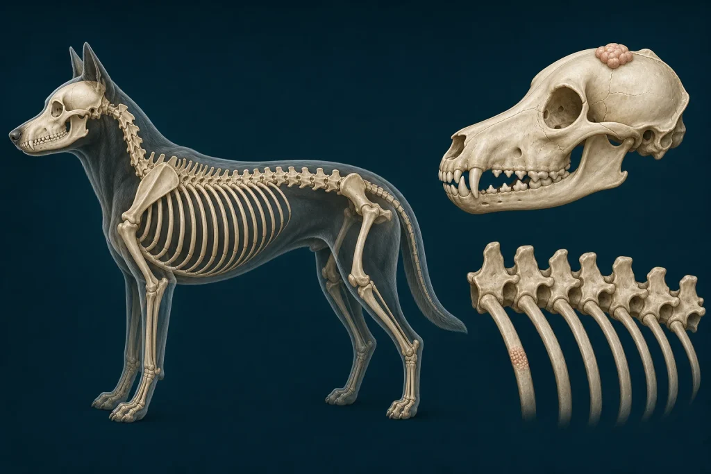
Overview
Multilobular Tumor of Bone is a nodular bone-producing tumor that most often arises from the flat bones of the skull. Many grow slowly, but continued expansion can compress the brain, eye, airway, or jaw and recurrence or metastasis risk varies with grade and completeness of removal.
Common signs
Owners may notice a firm immobile head lump, facial asymmetry, eye displacement, difficulty opening the mouth, chewing change, nasal signs, neurologic change, or pain from pressure. None of these signs is specific to multilobular tumor of bone, so a veterinary examination and appropriate testing are needed before naming the problem.
Common locations
The calvarium, maxilla, mandible, orbit, and other skull bones may be involved, and the visible contour may underestimate attachment or intracranial extension. Anatomy affects what can be sampled safely, whether a complete first surgery is realistic, and which nearby function needs protection.
Diagnosis discussion
Skull CT is commonly used to map bone involvement and plan biopsy or surgery. Histopathology supplies grade, while chest imaging and other staging are selected according to the confirmed diagnosis. The diagnostic plan should answer a decision: whether tissue type, local extent, stage, or patient health will change the next option. An imaging impression is not the same as a histologic diagnosis.
Urgency and when to contact a veterinarian
Arrange a prompt veterinary appointment for persistent or progressive signs. Seek urgent veterinary care for any of these signs: seizures, breathing difficulty, inability to eat or drink, sudden vision loss with pain, uncontrolled bleeding, or rapid neurologic decline. Call ahead when possible so the receiving team can prepare; do not delay emergency transport to take photographs or complete a log.
Treatment discussion
Wide planned surgery is the main local option when anatomy permits. Radiation or palliative care may be considered for incomplete removal, recurrence, or an unresectable site; reconstruction needs discussion before the first operation. The team should explain the goal of each option—diagnosis, local control, systemic control, symptom relief, or emergency stabilization—along with likely burdens, costs, travel, rechecks, and alternatives.
Home monitoring
Keep one dated home record of head measurements and photographs, eye position, jaw opening, chewing, swallowing, nasal airflow, pain, behavior, gait, and any new seizures. Use the same measurement or observation method each time and share trends with the veterinary team. Home tracking supports planned rechecks; it cannot determine grade, margins, or metastasis.
Questions to ask your veterinarian
- Does CT show intracranial, orbital, nasal, or jaw involvement?
- What biopsy track preserves the definitive surgical field?
- Can adequate margins and reconstruction be planned at the first operation?
- How will grade and margins determine imaging follow-up?