Canine Perivascular Wall Tumor: A Limb Mass May Extend Beyond What You Can Feel
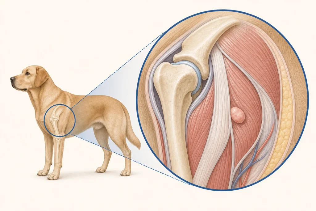
Overview
Perivascular Wall Tumor is a category of soft-tissue sarcoma arising from cells associated with blood-vessel walls. These tumors are often locally infiltrative and may recur after incomplete removal; microscopic diagnosis and grade matter more than an older label such as hemangiopericytoma alone.
Common signs
Owners may notice a slow-growing firm mass under the skin, commonly on a limb, with possible stretching, licking, ulceration, pain, or reduced motion as it enlarges. None of these signs is specific to perivascular wall tumor, so a veterinary examination and appropriate testing are needed before naming the problem.
Common locations
Distal and proximal limbs are frequent locations, but trunk and other subcutaneous sites occur; fascial planes and nearby neurovascular structures influence margins. Anatomy affects what can be sampled safely, whether a complete first surgery is realistic, and which nearby function needs protection.
Diagnosis discussion
Fine-needle aspiration may identify a mesenchymal pattern but can be inconclusive. Imaging for deep or fixed lesions, a strategically placed biopsy, and histopathology with grade and margins guide definitive planning. The diagnostic plan should answer a decision: whether tissue type, local extent, stage, or patient health will change the next option. An imaging impression is not the same as a histologic diagnosis.
Urgency and when to contact a veterinarian
Arrange a prompt veterinary appointment for persistent or progressive signs. Seek urgent veterinary care for any of these signs: uncontrolled bleeding, sudden severe pain or swelling, inability to use the limb, a rapidly opening wound, or collapse. Call ahead when possible so the receiving team can prepare; do not delay emergency transport to take photographs or complete a log.
Treatment discussion
Wide first-intent surgery is commonly discussed. Radiation may support local control after incomplete removal or at difficult sites, while systemic therapy is considered selectively based on grade, stage, and evidence. The team should explain the goal of each option—diagnosis, local control, systemic control, symptom relief, or emergency stabilization—along with likely burdens, costs, travel, rechecks, and alternatives.
Home monitoring
Keep one dated home record of two perpendicular measurements, photographs, skin mobility, ulceration, licking, limb use, joint range, pain, and any new nodules near the scar. Use the same measurement or observation method each time and share trends with the veterinary team. Home tracking supports planned rechecks; it cannot determine grade, margins, or metastasis.
Questions to ask your veterinarian
- Does imaging show extension along fascia, muscle, or a neurovascular bundle?
- Where should the biopsy tract be placed within the planned resection?
- What grade and margin language will the pathologist report?
- Would radiation consultation before re-excision preserve more options?