Canine Ureteral Tumors: A Rare Obstruction Between Kidney and Bladder
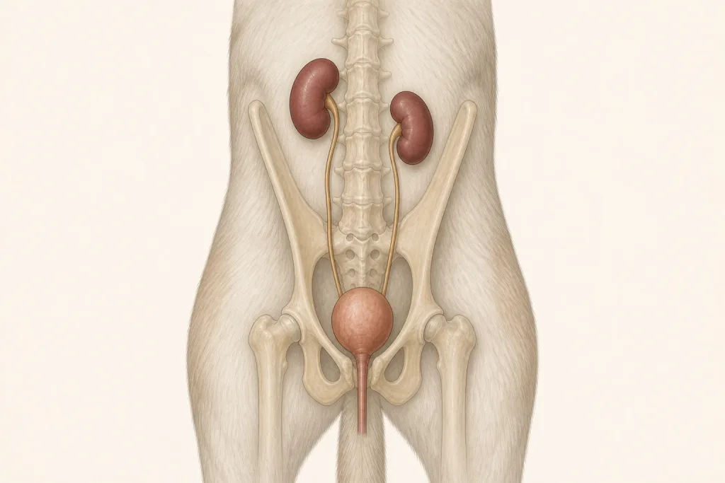
Overview
Ureteral Tumors are uncommon neoplasms affecting one of the narrow tubes that drain urine from a kidney to the bladder. A small lesion can obstruct flow and damage a kidney, but stones, strictures, inflammation, or compression from another mass are more common alternatives.
Common signs
Owners may notice abdominal or back pain, blood in urine, vomiting, appetite loss, lethargy, recurrent urinary infection, increased thirst, or no obvious signs despite kidney dilation on imaging. None of these signs is specific to ureteral tumors, so a veterinary examination and appropriate testing are needed before naming the problem.
Common locations
The lesion can occur near the kidney, along the abdominal ureter, or where it enters the bladder; unilateral obstruction may not stop visible urination. Anatomy affects what can be sampled safely, whether a complete first surgery is realistic, and which nearby function needs protection.
Diagnosis discussion
Kidney values, urinalysis and culture, expert ultrasound, CT urography, and endoscopic or surgical evaluation may be discussed. Imaging assesses obstruction, function of both kidneys, local spread, and other causes. The diagnostic plan should answer a decision: whether tissue type, local extent, stage, or patient health will change the next option. An imaging impression is not the same as a histologic diagnosis.
Urgency and when to contact a veterinarian
Arrange a prompt veterinary appointment for persistent or progressive signs. Seek urgent veterinary care for any of these signs: severe pain, repeated vomiting, collapse, inability to urinate, fever with marked weakness, or rapidly worsening kidney values. Call ahead when possible so the receiving team can prepare; do not delay emergency transport to take photographs or complete a log.
Treatment discussion
Surgical removal or reconstruction, ureteral stenting or bypass, kidney removal when necessary and safe, systemic therapy for selected histologies, and palliative management are individualized. The team should explain the goal of each option—diagnosis, local control, systemic control, symptom relief, or emergency stabilization—along with likely burdens, costs, travel, rechecks, and alternatives.
Home monitoring
Keep one dated home record of urine color and frequency, pain, appetite, vomiting, water intake, weight, fever if directed, and scheduled kidney-function and imaging rechecks. Use the same measurement or observation method each time and share trends with the veterinary team. Home tracking supports planned rechecks; it cannot determine grade, margins, or metastasis.
Questions to ask your veterinarian
- Is the obstruction unilateral, and how much function remains in each kidney?
- Could a stone, stricture, or external compression explain the imaging finding?
- Which procedure can restore drainage while preserving renal function?
- Is tissue diagnosis possible without delaying decompression?