Canine Hepatosplenic Lymphoma: Liver and Spleen Involvement Without Large Nodes
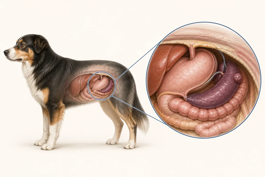
Overview
Hepatosplenic Lymphoma is a lymphoid cancer pattern centered in the liver and spleen, sometimes with little peripheral lymph-node enlargement. It can be aggressive and may involve blood or marrow, but infection, inflammation, other lymphoma forms, and histiocytic disease can produce overlapping organ changes.
Common signs
Owners may notice lethargy, reduced appetite, weight loss, vomiting, abdominal enlargement or discomfort, pale gums, fever, weakness, bruising, or abnormal liver and blood counts. None of these signs is specific to hepatosplenic lymphoma, so a veterinary examination and appropriate testing are needed before naming the problem.
Common locations
The spleen and liver are primary apparent sites, with potential marrow, blood, or other organ involvement despite normal-sized external nodes. Anatomy affects what can be sampled safely, whether a complete first surgery is realistic, and which nearby function needs protection.
Diagnosis discussion
CBC with smear, chemistry, coagulation assessment, abdominal imaging, carefully planned liver or spleen sampling, flow cytometry, clonality, marrow evaluation, and infectious-disease testing may be used. The diagnostic plan should answer a decision: whether tissue type, local extent, stage, or patient health will change the next option. An imaging impression is not the same as a histologic diagnosis.
Urgency and when to contact a veterinarian
Arrange a prompt veterinary appointment for persistent or progressive signs. Seek emergency veterinary care for any of these signs: collapse, very pale gums, severe abdominal pain or enlargement, uncontrolled bleeding, breathing difficulty, fever with profound weakness, or inability to stand. Call ahead when possible so the receiving team can prepare; do not delay emergency transport to take photographs or complete a log.
Treatment discussion
Systemic therapy and supportive care are considered once lineage and patient stability are clear. Surgery alone is generally not a complete strategy for diffuse lymphoid disease, though sampling or emergency splenic indications can alter the plan. The team should explain the goal of each option—diagnosis, local control, systemic control, symptom relief, or emergency stabilization—along with likely burdens, costs, travel, rechecks, and alternatives.
Home monitoring
Keep one dated home record of appetite, abdominal size, vomiting, gum color, bruising, fever if instructed, breathing, energy, and treatment side effects. Use the same measurement or observation method each time and share trends with the veterinary team. Home tracking supports planned rechecks; it cannot determine grade, margins, or metastasis.
Questions to ask your veterinarian
- Which sampling site offers the best balance of yield and bleeding risk?
- Do flow cytometry or clonality results define the lymphoid population?
- Is marrow or blood involvement present despite normal external nodes?
- What findings require stabilization before systemic therapy?