A Swollen Limb During Cancer Care: Measure the Change Without Massaging It
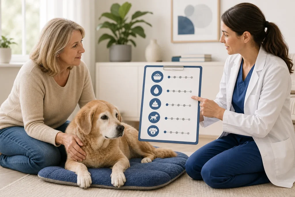
A limb can look larger because of edema, a focal mass, hematoma, inflammation, fluid leakage, or positioning. The safest home role is to document onset, distribution, pain, heat, color, function, and progression, then contact the care team. Treatment depends on the cause and tissue stability.
Why this deserves a plan
Lymphedema techniques are specialized and are not appropriate for every swollen limb. A tumor-weakened bone, fresh incision, infection, clot concern, bleeding tendency, or painful mass can change what pressure and movement are safe. Early assessment also protects skin before leakage or ulceration develops.
Prepare before the stressful moment
Ask whether the dog is at known risk after node removal, surgery, radiation, or pelvic or axillary disease. Keep an approved measuring location and method if the team recommends one. Use the same posture and time of day. Photograph the whole limb and a close view without squeezing tissue.
What to observe and record
Record where swelling begins and ends, circumference at the marked location, pitting only if the clinician taught you to assess it, temperature difference, color, skin leakage, pain, gait, breathing, and medication timing. Note whether the change is sudden or gradual and whether more than one limb is involved.
When to call or escalate
Sudden severe swelling, marked pain, cold or discolored paw, bleeding, fever with illness, breathing difficulty, collapse, or rapid loss of function needs urgent assessment. New skin breakdown, fluid leakage, or progressive swelling deserves prompt contact even when the dog remains bright.
What not to improvise
Do not massage, wrap tightly, apply heat, use a compression garment, or increase exercise without clearance. Do not assume all swelling after surgery is normal. Human diuretics or topical products can be harmful and may delay identification of obstruction, infection, or hemorrhage.
Describe the geography of the swelling
Instead of reporting only that the limb is “bigger,” note whether the change begins near the toes, surrounds a joint, follows an incision, sits over a known mass, or involves the entire limb. Compare skin color, warmth, comfort, paw placement, and any fluid leakage without pressing repeatedly. This distribution helps the care team decide which causes and examinations need priority.
If the clinic requests circumference tracking, have staff mark the exact landmark and demonstrate tape placement and limb position. Measurements taken at different locations can create a false trend, while repeated tight handling can hurt fragile tissue. Record the value with time, posture, activity, medication, and a photograph from the same angle. A sudden functional or skin change should prompt contact even if the tape measurement appears similar. Also note whether collars, bandages, or harness straps cross the swollen area during movement or rest.
Take these questions to the care team
- What causes must be ruled out first?
- Is measurement useful and where?
- Is compression or manual therapy cleared?
- How should skin be protected?
- Which change requires emergency care?
Medical disclaimer: This article is educational and cannot diagnose, treat, or determine prognosis for an individual dog. It does not replace an examination or an individualized plan from a licensed veterinarian.
Sources and further reading
- American Association of Rehabilitation Veterinarians: Model Standards
- AAHA: Supportive and Symptomatic Care
- AAHA: 2022 Pain Management Guidelines
- American College of Veterinary Surgeons: Postoperative Care
