Canine Conjunctival Tumors: A Persistent Eye-Surface Mass Needs Ophthalmic Assessment
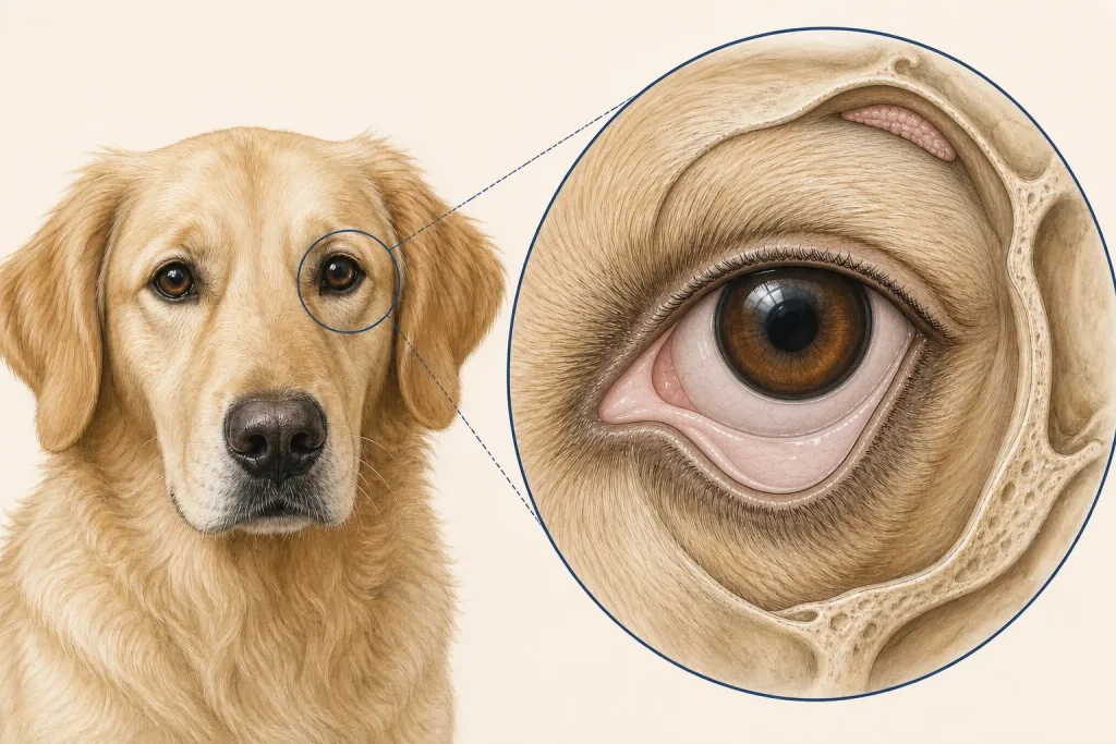
Overview
Conjunctival Tumors are a varied group of growths arising from the membrane lining the eyelids and covering part of the eye. Benign and malignant lesions, inflammation, foreign-body reactions, and prolapsed tissue can look similar, while even a small mass may irritate or damage the cornea.
Common signs
Owners may notice a pink, red, pigmented, or irregular mass; persistent redness; discharge; squinting; rubbing; bleeding; corneal cloudiness; or apparent growth from the inner eyelid. None of these signs is specific to conjunctival tumors, so a veterinary examination and appropriate testing are needed before naming the problem.
Common locations
Bulbar, palpebral, and third-eyelid conjunctiva are distinct sites; the lesion’s attachment and extension toward the orbit or cornea affect treatment. Anatomy affects what can be sampled safely, whether a complete first surgery is realistic, and which nearby function needs protection.
Diagnosis discussion
A complete ophthalmic examination, fluorescein assessment when appropriate, eye-pressure testing, photography, imaging for deeper lesions, and biopsy or excision with histopathology may be discussed. The diagnostic plan should answer a decision: whether tissue type, local extent, stage, or patient health will change the next option. An imaging impression is not the same as a histologic diagnosis.
Urgency and when to contact a veterinarian
Arrange a prompt veterinary appointment for persistent or progressive signs. Seek urgent veterinary care for any of these signs: sudden severe eye pain, a blue or white cloudy cornea, inability to open the eye, rapid swelling, heavy bleeding, trauma, or sudden vision loss. Call ahead when possible so the receiving team can prepare; do not delay emergency transport to take photographs or complete a log.
Treatment discussion
Local excision, cryotherapy or other adjuncts, wider surgery, radiation, systemic therapy for selected histologies, or monitoring may be considered. Preserving comfort and vision must be balanced with complete diagnosis and local control. The team should explain the goal of each option—diagnosis, local control, systemic control, symptom relief, or emergency stabilization—along with likely burdens, costs, travel, rechecks, and alternatives.
Home monitoring
Keep one dated home record of mass size and color, squinting, rubbing, discharge, bleeding, corneal clarity, vision behavior, appetite, and response to prescribed eye medication. Use the same measurement or observation method each time and share trends with the veterinary team. Home tracking supports planned rechecks; it cannot determine grade, margins, or metastasis.
Questions to ask your veterinarian
- Which conjunctival surface is the true site of origin?
- Is the cornea, third eyelid, orbit, or globe involved?
- Can excision provide both diagnosis and adequate margins without compromising function?
- What postoperative sign suggests corneal injury or pressure change?