Canine Gastrointestinal Stromal Tumors: A Mesenchymal GI Cancer
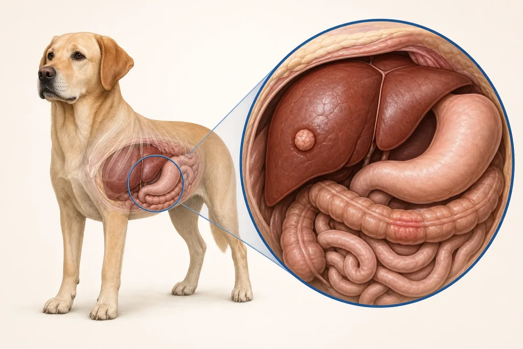
Overview
Gastrointestinal stromal tumors, often shortened to GIST, are mesenchymal tumors of the gastrointestinal tract. They can resemble other spindle-cell tumors, so histopathology and immunohistochemical markers are important for accurate classification.
Common signs
Vomiting, diarrhea, dark or bloody stool, anemia, reduced appetite, weight loss, abdominal discomfort, weakness, or an abdominal mass may occur. Some lesions are discovered during imaging or surgery for another concern.
Common locations
GIST can arise along the stomach or intestines, with the large intestine and cecal region reported sites in dogs. Local growth, bleeding, obstruction, or spread to liver or peritoneum may influence presentation.
Diagnosis discussion
Bloodwork can reveal anemia or systemic effects, while ultrasound or CT helps define the mass and plan surgery. Cytology may be nondiagnostic. Histopathology plus immunohistochemistry, including markers selected by the pathologist, distinguishes GIST from other sarcomas.
Urgency and when to contact a veterinarian
Collapse, very pale gums, repeated unproductive vomiting, a swollen painful abdomen, black tarry stool with weakness, or inability to pass stool may represent bleeding or obstruction and needs urgent care.
Treatment discussion
Surgical removal is commonly considered for localized disease. Pathology, margins, mitotic activity, spread, and molecular features may lead to oncology discussion of targeted or systemic therapy. Supportive care addresses bleeding, nausea, pain, and nutrition.
Home monitoring
Track appetite, vomiting, stool color and frequency, abdominal size or pain, weight, gum color, energy, and medication tolerance. Photograph stool only if useful to the clinic and do not delay care for suspected bleeding.
Questions to ask your veterinarian
- Will pathology use immunohistochemistry to distinguish GIST from other sarcomas?
- Does imaging suggest obstruction, bleeding, or spread?
- Can the mass be removed with acceptable bowel margins?
- Which pathology or molecular results would justify a targeted-therapy discussion?