Canine Splenic Mass: A Tissue Diagnosis Matters Before Naming the Tumor
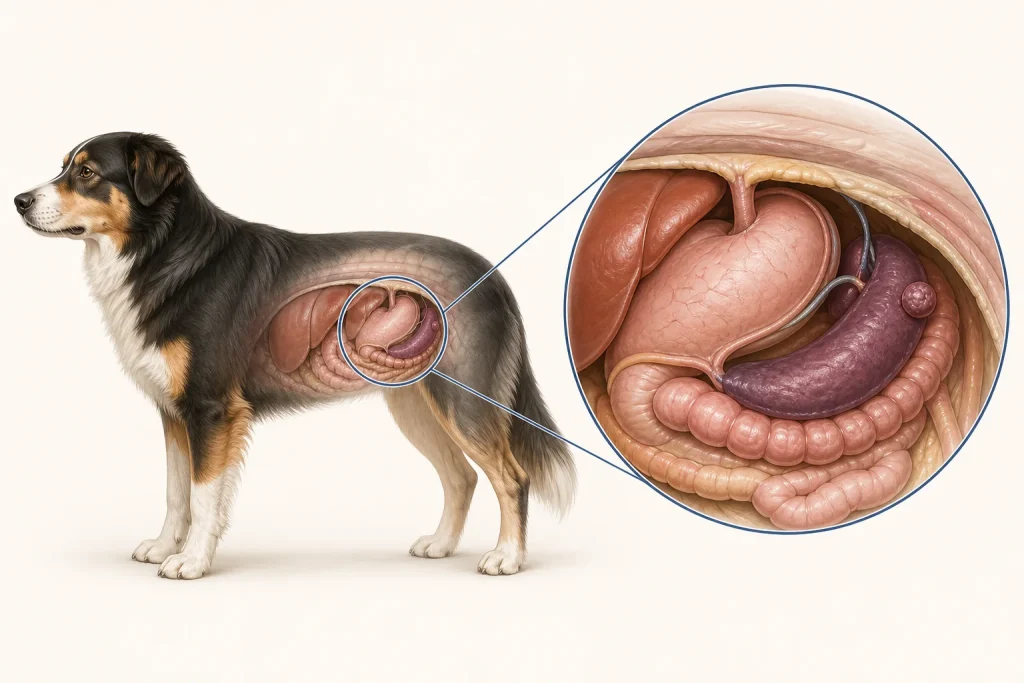
Overview
Splenic mass is an anatomic description that includes benign and malignant causes. Imaging can estimate size, bleeding and other lesions but usually cannot prove histologic type.
Common signs
No signs when found incidentally, or reduced appetite, intermittent weakness, abdominal enlargement, pale gums, collapse and rapid breathing if bleeding occurs.
Common locations
Within the spleen; free abdominal fluid or lesions in the liver, omentum or other organs may be evaluated concurrently.
Diagnosis discussion
Assessment commonly includes blood count, chemistry, coagulation testing, abdominal ultrasound and chest imaging, with heart ultrasound in selected cases. Cytology is not always safe or diagnostic. Surgical removal followed by histopathology often provides the definitive diagnosis.
Urgency and when to contact a veterinarian
Any collapse, pale gums, marked weakness, rapid breathing or sudden abdominal distension requires immediate emergency care. A stable incidental mass still needs a timely plan because bleeding risk and tumor type are uncertain.
Treatment discussion
The team may discuss monitoring in selected stable cases, splenectomy for diagnosis and prevention or control of bleeding, staging, and subsequent treatment based on pathology. Surgical candidacy depends on stability, other disease, detectable spread and family goals.
Home monitoring
Until a plan is set, avoid rough activity and know the nearest emergency facility. Record appetite, energy, gum color only if your veterinarian teaches you, resting breathing and abdominal appearance. Do not treat a suspected bleed with home remedies.
Questions to ask your veterinarian
- Is there evidence of active or recent bleeding right now?
- Which findings make a benign or malignant cause more likely, and what remains uncertain?
- What are this dog's anesthesia and surgery risks?
- Will every removed tissue be submitted for histopathology?