Canine Cutaneous Hemangioma and Hemangiosarcoma: Vascular Skin Masses Need Tissue Context
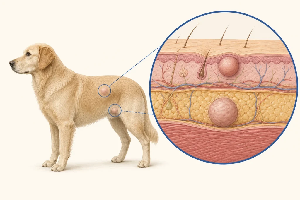
Overview
Hemangioma and hemangiosarcoma arise from vascular tissue. Superficial sun-associated lesions can appear as red, purple or black spots or nodules, while deeper subcutaneous vascular tumors may behave more aggressively; bruising and other tumors can look alike.
Common signs
Red to dark-purple spots, plaques or nodules, easy bleeding, crusting, bruised appearance or recurrent scabbing. Some lesions are painless until traumatized.
Common locations
Sparsely haired, sun-exposed ventral skin is a recognized setting for superficial lesions; vascular tumors can also occur in deeper skin or subcutaneous tissue.
Diagnosis discussion
Cytology may be nondiagnostic when a lesion yields mostly blood. Biopsy or complete excision with histopathology establishes type and depth. The veterinarian may recommend staging for malignant or deeper disease.
Urgency and when to contact a veterinarian
Promptly assess a recurrently bleeding or bruise-like mass. Emergency care is needed for uncontrolled bleeding, pale gums, weakness, collapse or breathing difficulty.
Treatment discussion
Surgical removal is a common treatment for localized skin lesions. Histologic type, depth, completeness and multiplicity determine whether surveillance, staging or additional therapy should be discussed.
Home monitoring
Prevent licking and trauma, use sun-exposure reduction recommended by the veterinarian, and record new spots, bleeding episodes, bruising, energy and gum color.
Questions to ask your veterinarian
- Could this lesion be safely sampled, or is planned excision more informative?
- Does pathology distinguish superficial from deeper aggressive disease?
- Is staging needed based on depth and histology?
- What is the home plan for a bleeding episode?