Canine Myxosarcoma: A Soft-Tissue Mass With High Local-Recurrence Concern
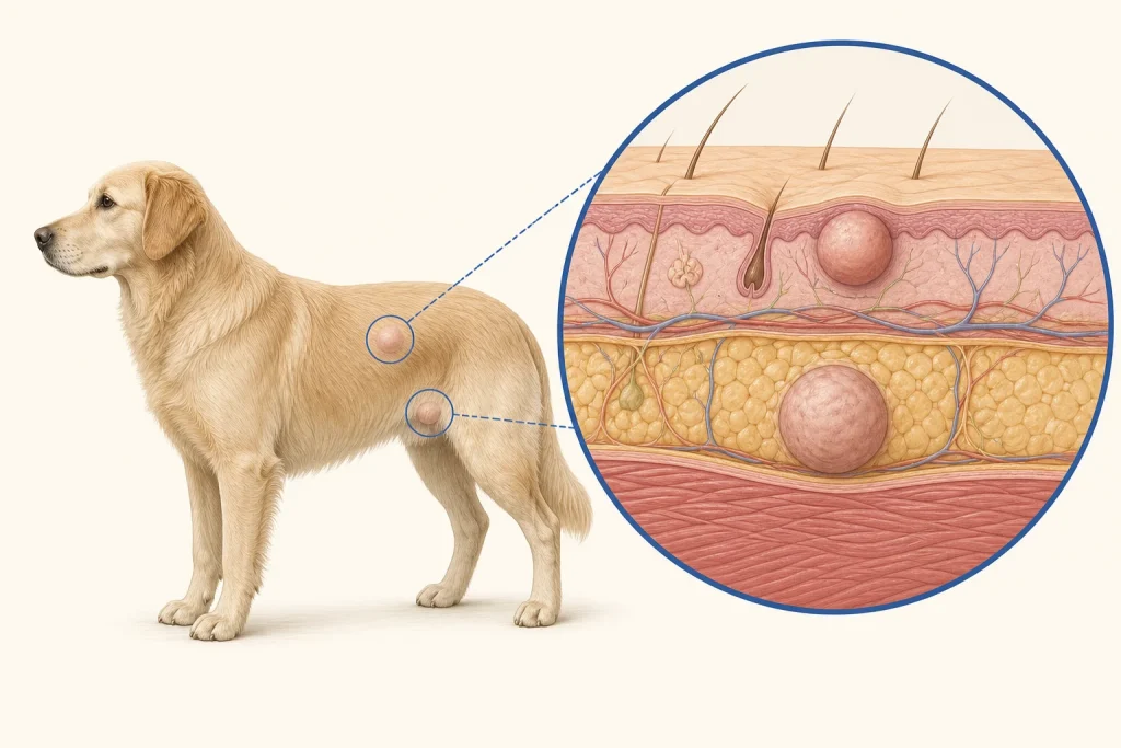
Overview
Myxosarcoma is a malignant soft-tissue tumor whose cells produce a gelatinous matrix. It may be soft or poorly defined and is known chiefly for local infiltration and recurrence risk rather than a predictable appearance.
Common signs
A slow-growing soft, fluctuant or firm mass under the skin or within deeper tissue. Larger lesions may stretch skin, limit movement, become painful or ulcerate.
Common locations
Trunk and limbs are common, but the tumor can arise in other connective tissues. Proximity to joints, nerves and body-wall structures affects surgical options.
Diagnosis discussion
Fine-needle samples may yield little material. Core or incisional biopsy and imaging can be more informative before definitive surgery. Histopathology confirms subtype, grade and margin status.
Urgency and when to contact a veterinarian
Promptly evaluate a persistent or enlarging mass. Seek urgent care if it rapidly impairs walking, becomes acutely painful, bleeds heavily or compromises breathing or elimination.
Treatment discussion
Wide surgical excision is often the central local-control discussion. Radiation may be considered when complete margins are not feasible; other therapy and staging depend on grade and patient factors.
Home monitoring
Use fixed landmarks for measurements and record texture, mobility, gait and skin condition. After surgery, photograph the scar and report any new thickening or nodule early.
Questions to ask your veterinarian
- Would biopsy before surgery improve margin planning?
- Should CT or MRI map the lesion's deep extent?
- What is the expected local-recurrence risk from grade and margins?
- Would radiation meaningfully improve local control in this case?