Canine Oral Squamous Cell Carcinoma: Persistent Mouth Lesions Need Biopsy
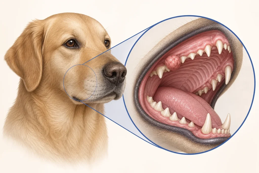
Overview
Oral squamous cell carcinoma arises from lining cells of the mouth. It may resemble dental infection, gum inflammation or a nonhealing ulcer; location within the mouth influences local invasion, metastatic risk and treatment options.
Common signs
Bad breath, drooling, oral bleeding, a visible gum or tongue mass, loose teeth, facial swelling, chewing on one side, dropping food or weight loss.
Common locations
Gums, tonsil, tongue and other oral surfaces. Tumors at different sites can have importantly different behavior, so the pathology report should be interpreted with exact location.
Diagnosis discussion
A thorough oral and dental examination plus biopsy establishes the diagnosis. Dental radiographs or CT define bone involvement; regional lymph nodes may be sampled even when not obviously enlarged, with chest imaging selected for staging.
Urgency and when to contact a veterinarian
Promptly assess any oral mass, unexplained loose tooth or ulcer that persists. Urgent care is needed for uncontrolled bleeding, breathing difficulty, inability to swallow, dehydration or severe pain.
Treatment discussion
Wide oral surgery is often the main local-control option when feasible. Radiation and selected systemic therapies may be discussed according to site, margins, nodes and stage; nutrition and pain control belong in the plan from the start.
Home monitoring
Record meal duration, food texture tolerated, drooling, bleeding, breath odor, weight and pain behavior. Do not force the mouth open or disturb a friable lesion.
Questions to ask your veterinarian
- Could dental disease and tumor coexist, and which area should be biopsied?
- Would CT change the surgical or radiation plan?
- Should both regional lymph nodes be sampled?
- What is the feeding and pain plan before and after treatment?