Canine Peripheral Odontogenic Fibroma: A Common Epulis-Type Mass Needs Pathology
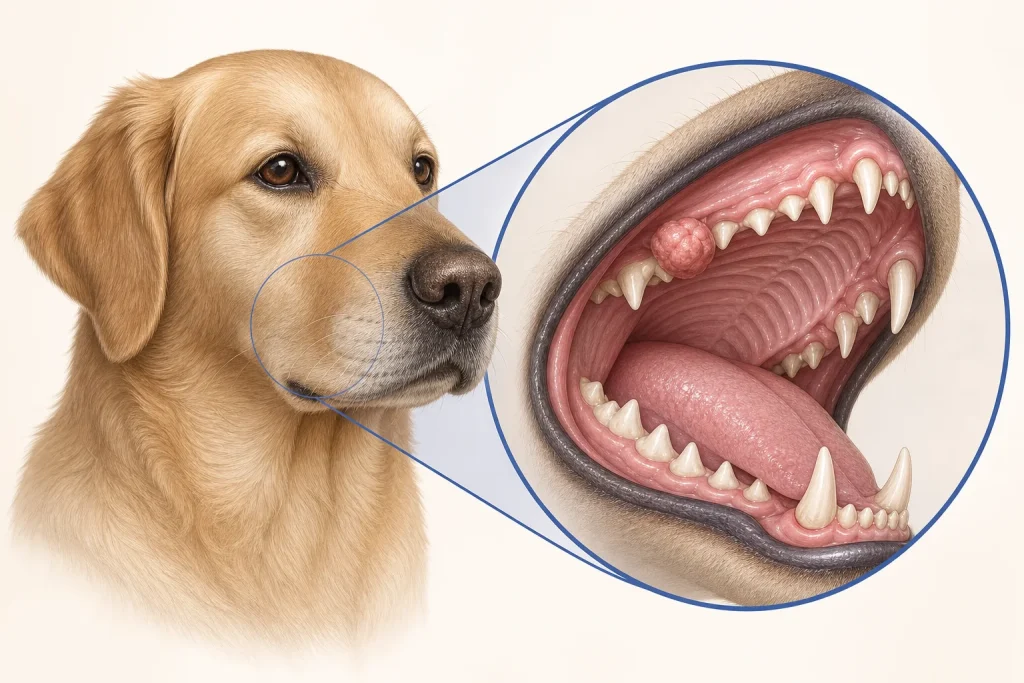
Overview
Peripheral odontogenic fibroma is a generally benign gum tumor historically included under the vague term epulis. Other benign and malignant oral tumors share that appearance, so pathology is more useful than the descriptive nickname.
Common signs
A firm smooth or lobulated gum mass, food trapping, bad breath, minor bleeding, tooth displacement or chewing discomfort. Small lesions may be found during routine dental examination.
Common locations
Gingiva adjacent to teeth in the upper or lower jaw. The relationship to periodontal tissues and bone affects removal.
Diagnosis discussion
Oral examination and dental radiographs assess the associated tooth and bone. Biopsy or complete excision with histopathology confirms the tumor and distinguishes it from more invasive odontogenic and malignant lesions.
Urgency and when to contact a veterinarian
Arrange prompt evaluation for any new gum mass. Earlier care is needed if it grows quickly, bleeds repeatedly, causes pain, prevents eating or is accompanied by facial swelling.
Treatment discussion
Complete surgical removal, sometimes including involved periodontal tissue or tooth management, is commonly curative. Incomplete removal can allow local recurrence, so pathology and recheck matter.
Home monitoring
Photograph only when safe, record chewing comfort, breath odor, bleeding and food intake, and follow postoperative soft-food and oral-hygiene instructions.
Questions to ask your veterinarian
- Can this be removed completely at the time of biopsy?
- Do dental radiographs show bone or tooth involvement?
- Will the specimen receive a specific histopathologic name and margin assessment?
- What local change suggests recurrence?