Canine Acanthomatous Ameloblastoma: Benign Histology, Locally Destructive Behavior
Overview
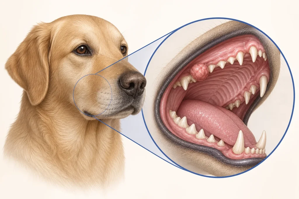
Canine acanthomatous ameloblastoma is an odontogenic tumor that does not usually spread to distant organs but can invade jaw bone extensively. Its locally aggressive behavior means that casual removal of the visible portion is rarely an adequate plan.
Common signs
A firm gum mass, displaced or loose teeth, oral bleeding, bad breath, drooling, facial swelling or difficulty chewing. Some are discovered during dental care.
Common locations
Usually the tooth-bearing lower or upper jaw, often near the front of the mouth, with extension into adjacent bone.
Diagnosis discussion
Biopsy establishes the odontogenic diagnosis. Dental radiographs and especially CT can map bone involvement and define the true treatment field before definitive surgery or radiation planning.
Urgency and when to contact a veterinarian
Promptly evaluate any persistent gum growth. Urgent care is appropriate for significant bleeding, severe pain, inability to eat or drink, or breathing compromise.
Treatment discussion
Jaw surgery with appropriately planned margins is a common route to durable local control. Radiation may be considered in selected cases where surgery is not feasible or accepted; repeated marginal debulking carries recurrence risk.
Home monitoring
Record chewing comfort, food intake, drooling, bleeding, weight and facial change. Follow oral-care and diet instructions after treatment and attend long-term local rechecks.
Questions to ask your veterinarian
- Does biopsy confirm an odontogenic tumor rather than another oral cancer?
- How far into jaw bone does CT show extension?
- What function should we expect after the proposed jaw surgery?
- What local-recurrence monitoring is needed long term?