Canine Hepatocellular Carcinoma: Liver Mass Pattern and Resectability Matter
Overview
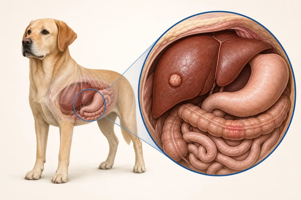
Hepatocellular carcinoma is a malignant tumor of liver cells. A single large or massive lesion may be surgically removable and behave differently from multifocal or diffuse liver disease, so imaging pattern and anatomic location are central to planning.
Common signs
Reduced appetite, weight loss, lethargy, vomiting, increased thirst, abdominal enlargement, jaundice or weakness; many masses are discovered during imaging for another reason.
Common locations
Any liver lobe, as a solitary massive lesion, multiple nodules or diffuse involvement. Relationship to major vessels and bile ducts affects surgery.
Diagnosis discussion
Bloodwork assesses liver function, anemia, clotting and anesthesia risk but does not name the mass. Ultrasound and CT define distribution and resectability. Cytology or biopsy may be considered, while surgical histopathology often provides the final diagnosis.
Urgency and when to contact a veterinarian
Promptly investigate a liver mass or persistent liver-related signs. Collapse, pale gums, a rapidly distended abdomen, uncontrolled bleeding, severe weakness or marked jaundice warrants urgent care.
Treatment discussion
Liver-lobe surgery is commonly discussed for a solitary resectable mass. Multifocal, diffuse or metastatic disease may prompt systemic, interventional or palliative options; pathology and stage guide follow-up.
Home monitoring
Track appetite, weight, vomiting, abdominal size, gum color, jaundice, energy and medication effects. Follow any activity restriction if bleeding risk is suspected.
Questions to ask your veterinarian
- Is the imaging pattern solitary, nodular or diffuse?
- Would CT materially change surgical planning?
- How are clotting and bleeding risk being assessed?
- Which pathology findings guide oncology follow-up?