Canine Exocrine Pancreatic Tumors: Vague Signs, Careful Imaging
Overview
Tumors arising from the exocrine pancreas are rare in dogs and may be difficult to detect early. Pancreatitis and diseases of the liver, gallbladder, stomach, or intestine can produce similar signs, while imaging may not always distinguish inflammation from neoplasia.
Common signs
Reduced appetite, vomiting, weight loss, abdominal discomfort, lethargy, diarrhea, jaundice, or an abdominal mass may occur. Signs are often nonspecific and can fluctuate.
Common locations
The pancreas lies beside the stomach and small intestine and near the liver, bile duct, and major vessels. Local invasion or spread to liver, lymph nodes, peritoneum, or other sites may affect options.
Diagnosis discussion
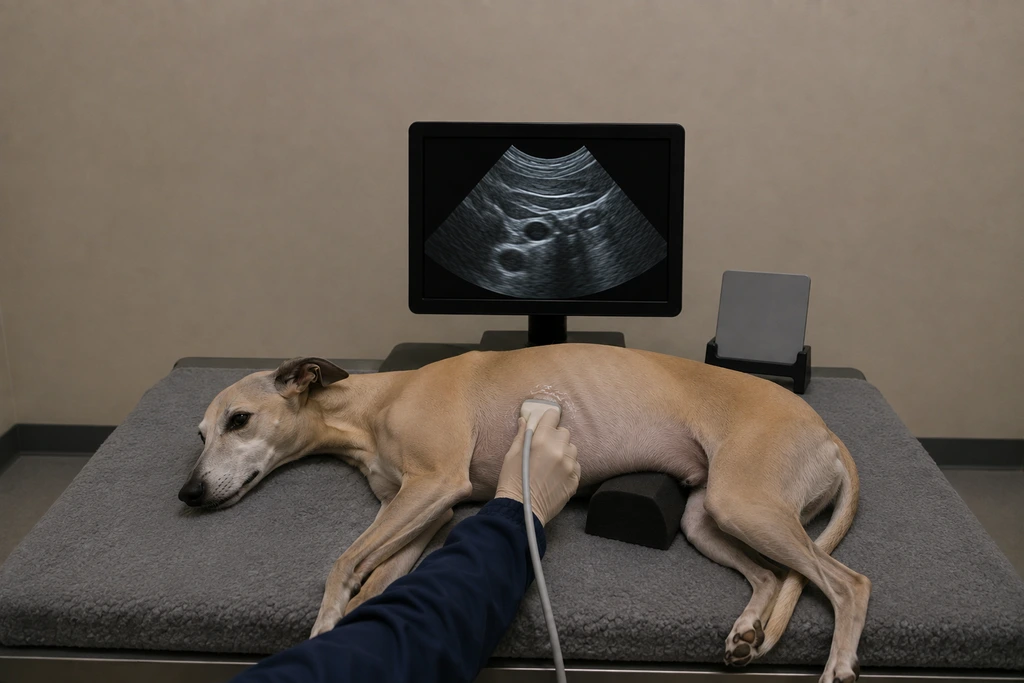
The veterinarian may use bloodwork, abdominal ultrasound or CT, and selective cytology or biopsy. Pancreatic enzyme tests address pancreatitis but do not by themselves diagnose or exclude a tumor. Histopathology provides the most specific classification when tissue can be obtained safely.
Urgency and when to contact a veterinarian
Repeated vomiting, inability to keep water down, yellow gums or eyes, severe abdominal pain, a distended abdomen, collapse, or profound weakness needs urgent assessment.
Treatment discussion
Surgery may be discussed for a localized resectable lesion; systemic treatment or palliative support may be considered for invasive or metastatic disease. Nausea, nutrition, hydration, pain, and bile-flow complications require active supportive planning.
Home monitoring
Track meals offered and eaten, vomiting, stool, water intake, abdominal comfort, weight, jaundice, energy, and all medication effects. Report a rapid decline rather than waiting for a planned scan.
Questions to ask your veterinarian
- Which noncancer pancreatic or biliary conditions remain possible?
- Would CT or another imaging study change surgical planning?
- Can tissue be obtained safely and will it change treatment?
- What nausea, pain, and nutrition plan should start now?