CT and MRI: Cross-Sectional Imaging for Tumor Decisions
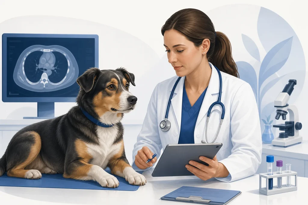
Quick summary
CT and MRI create cross-sectional datasets that can map tumors beyond what is visible or palpable. CT uses x-rays and is fast and strong for many chest, bone, nasal, and surgical-planning questions; MRI offers superior soft-tissue contrast and is commonly used for brain, spinal, nerve, and selected soft-tissue disease.
What it is
CT acquires x-ray attenuation data, often before and after intravenous contrast. MRI uses a strong magnetic field and radiofrequency signals. Both require precise positioning; MRI generally takes longer, and veterinary patients commonly require sedation or general anesthesia.
How it is discussed in tumor care
They may define local extent, vessel or bone involvement, surgical margins, radiation fields, neurologic lesion location, chest disease, or targets for biopsy. The best modality follows the clinical question rather than the idea that one scan is universally “more advanced.”
Potential role
A radiologist’s report integrates imaging with the history and can recommend another modality or sampling. Images can also be transferred in DICOM format for referral planning or second review.
Typical use context
CT is often considered for nasal, oral, lung, bone, adrenal, chest, and complex surgical cases. MRI is often considered for brain, spinal cord, nerve roots, and soft-tissue contrast questions.
Evidence snapshot
CT and MRI are established modalities directed and interpreted by veterinary imaging specialists. Their diagnostic accuracy depends on protocol, equipment, contrast use, motion control, lesion type, and the question asked.
Safety notes
Anesthesia or sedation, contrast reactions, kidney or cardiovascular considerations, radiation exposure from CT, and MRI magnet safety require screening. Facilities give case-specific fasting and medication instructions.
Interactions / cautions
Imaging phenotype is not always histologic diagnosis and cannot prove absence of microscopic spread. Keep the radiology report plus original DICOM files, not screenshots alone.
Questions to ask your veterinarian
- Which modality best answers our specific anatomical question?
- Will contrast and anesthesia be used, and what screening is needed?
- Will the scan guide biopsy, surgery, or radiation planning?
- Can we receive the report and DICOM images for referral?