Canine Tonsillar Squamous Cell Carcinoma: Subtle Throat Signs Can Mask Advanced Disease
Overview
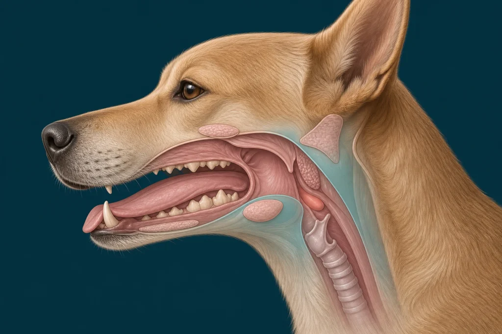
Tonsillar Squamous Cell Carcinoma is a malignant epithelial cancer arising from tonsillar tissue at the back of the mouth. The primary lesion can be difficult to see during an awake examination and may spread to regional lymph nodes, so a small visible mass does not define the stage.
Common signs
Owners may notice repeated swallowing, gagging, cough-like throat clearing, drooling, bad breath, reluctance to eat firm food, blood-tinged saliva, weight loss, or a new lump high in the neck. None of these signs is specific to tonsillar squamous cell carcinoma, so a veterinary examination and appropriate testing are needed before naming the problem.
Common locations
The palatine tonsils sit deep in the oropharynx near important swallowing structures and lymphatic drainage pathways. Anatomy affects what can be sampled safely, whether a complete first surgery is realistic, and which nearby function needs protection.
Diagnosis discussion
A careful sedated oral examination, cross-sectional imaging, biopsy, regional lymph-node sampling, and chest staging may be proposed. Histopathology is needed because inflammation and other oral tumors can create similar signs. The diagnostic plan should answer a decision: whether tissue type, local extent, stage, or patient health will change the next option. An imaging impression is not the same as a histologic diagnosis.
Urgency and when to contact a veterinarian
Arrange a prompt veterinary appointment for persistent or progressive signs. Seek emergency veterinary care for any of these signs: labored or noisy breathing at rest, inability to swallow, repeated choking, significant oral bleeding, collapse, or sudden weakness. Call ahead when possible so the receiving team can prepare; do not delay emergency transport to take photographs or complete a log.
Treatment discussion
Treatment may combine surgery, radiation, and systemic therapy according to local extent, lymph-node findings, distant spread, and the dog’s ability to eat and breathe comfortably. Supportive nutrition and pain control are part of the plan, not an afterthought. The team should explain the goal of each option—diagnosis, local control, systemic control, symptom relief, or emergency stabilization—along with likely burdens, costs, travel, rechecks, and alternatives.
Home monitoring
Keep one dated home record of swallow count, gagging, cough, drooling, oral odor, food texture tolerated, meal completion, weight, neck swelling, breathing sounds, and pain behaviors. Use the same measurement or observation method each time and share trends with the veterinary team. Home tracking supports planned rechecks; it cannot determine grade, margins, or metastasis.
Questions to ask your veterinarian
- Has each tonsil and the full oropharynx been examined under appropriate sedation?
- Which lymph nodes should be sampled for staging?
- Would radiation planning require CT before biopsy or surgery?
- How will nutrition and airway safety be supported during treatment?