Canine Tongue Tumors: Persistent Ulcers, Thickening, or Bleeding Need Biopsy
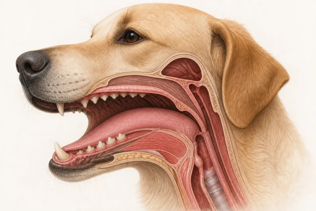
Overview
Tongue Tumors are a mixed group of epithelial, melanocytic, mesenchymal, and other tumors that develop within the tongue. Trauma, dental contact, infection, and immune disease can look similar, while tumor behavior differs markedly by histology and exact location.
Common signs
Owners may notice a nonhealing sore, focal thickening, a raised or pigmented mass, bleeding, drooling, pawing at the mouth, dropping food, altered tongue movement, bad breath, or reduced interest in eating. None of these signs is specific to tongue tumors, so a veterinary examination and appropriate testing are needed before naming the problem.
Common locations
Lesions may affect the mobile front of the tongue, its lateral edges, or the less visible base near the throat; location changes both function and surgical options. Anatomy affects what can be sampled safely, whether a complete first surgery is realistic, and which nearby function needs protection.
Diagnosis discussion
The team may recommend a sedated oral examination, imaging for deep or caudal lesions, biopsy, lymph-node assessment, and staging selected for the histologic diagnosis. A photograph cannot distinguish tumor from ulceration or inflammation. The diagnostic plan should answer a decision: whether tissue type, local extent, stage, or patient health will change the next option. An imaging impression is not the same as a histologic diagnosis.
Urgency and when to contact a veterinarian
Arrange a prompt veterinary appointment for persistent or progressive signs. Seek urgent veterinary care for any of these signs: uncontrolled bleeding, breathing difficulty, inability to take water, repeated aspiration-like coughing, collapse, or rapidly worsening swelling. Call ahead when possible so the receiving team can prepare; do not delay emergency transport to take photographs or complete a log.
Treatment discussion
Surgery may be appropriate for selected localized lesions, with reconstruction and feeding planning tailored to the amount and part of tongue involved. Radiation, systemic therapy, or comfort-focused care may be discussed for other tumor types or stages. The team should explain the goal of each option—diagnosis, local control, systemic control, symptom relief, or emergency stabilization—along with likely burdens, costs, travel, rechecks, and alternatives.
Home monitoring
Keep one dated home record of bleeding episodes, drooling, tongue mobility, meal duration, food dropped, water intake, coughing during meals, oral odor, body weight, and pain when yawning or chewing. Use the same measurement or observation method each time and share trends with the veterinary team. Home tracking supports planned rechecks; it cannot determine grade, margins, or metastasis.
Questions to ask your veterinarian
- Does the lesion extend deeper than its visible surface?
- What biopsy approach best preserves a later definitive procedure?
- How might the proposed treatment affect eating, drinking, and tongue function?
- What feeding or airway signs should trigger same-day contact?