Canine Mesothelioma: Recurrent Chest or Abdominal Fluid Needs a Cause
Overview
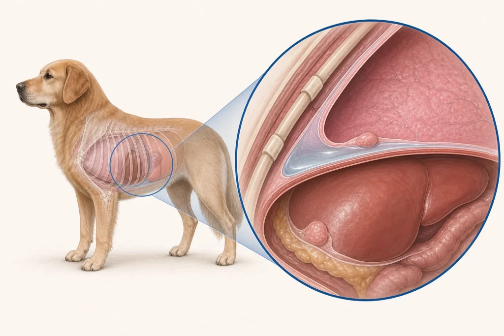
Mesothelioma is a malignant tumor of the thin lining of the chest, abdomen, heart sac, or more than one body cavity. Diffuse surface disease may be hard to see on routine imaging, and fluid cytology is often nondiagnostic because reactive lining cells can resemble tumor cells.
Common signs
Owners may notice rapid or labored breathing, abdominal enlargement, reduced stamina, cough, appetite loss, weight loss, weakness, or recurrence after fluid drainage. None of these signs is specific to mesothelioma, so a veterinary examination and appropriate testing are needed before naming the problem.
Common locations
Pleura, peritoneum, and pericardium are the main serosal surfaces; disease can be multifocal rather than a single removable mass. Anatomy affects what can be sampled safely, whether a complete first surgery is realistic, and which nearby function needs protection.
Diagnosis discussion
Fluid analysis, ultrasound, CT, thoracoscopy or laparoscopy, targeted biopsy, and histopathology with immunohistochemistry may be discussed. The team must also investigate infection, heart disease, bleeding, and other cancers that cause effusion. The diagnostic plan should answer a decision: whether tissue type, local extent, stage, or patient health will change the next option. An imaging impression is not the same as a histologic diagnosis.
Urgency and when to contact a veterinarian
Arrange a prompt veterinary appointment for persistent or progressive signs. Seek emergency veterinary care for any of these signs: breathing difficulty, blue or pale gums, collapse, inability to lie comfortably, a rapidly enlarging abdomen, or profound weakness. Call ahead when possible so the receiving team can prepare; do not delay emergency transport to take photographs or complete a log.
Treatment discussion
Treatment may focus on controlling fluid, systemic or intracavitary therapy in selected cases, surgery for limited components, and palliative care. Decisions are based on confirmed diagnosis, affected cavity, recurrence rate, breathing, and comfort. The team should explain the goal of each option—diagnosis, local control, systemic control, symptom relief, or emergency stabilization—along with likely burdens, costs, travel, rechecks, and alternatives.
Home monitoring
Keep one dated home record of sleeping respiratory rate, breathing effort, abdominal girth, appetite, mobility, sleep, fluid-drainage dates, and the interval until signs return. Use the same measurement or observation method each time and share trends with the veterinary team. Home tracking supports planned rechecks; it cannot determine grade, margins, or metastasis.
Questions to ask your veterinarian
- Which noncancer causes of the effusion have been excluded?
- Will fluid cytology answer the question, or is a targeted tissue biopsy needed?
- Which cavity is driving current symptoms and treatment priorities?
- How will we recognize and respond to fluid recurrence at home?