Canine Mediastinal Lymphoma: A Chest Mass Can Create an Airway Emergency
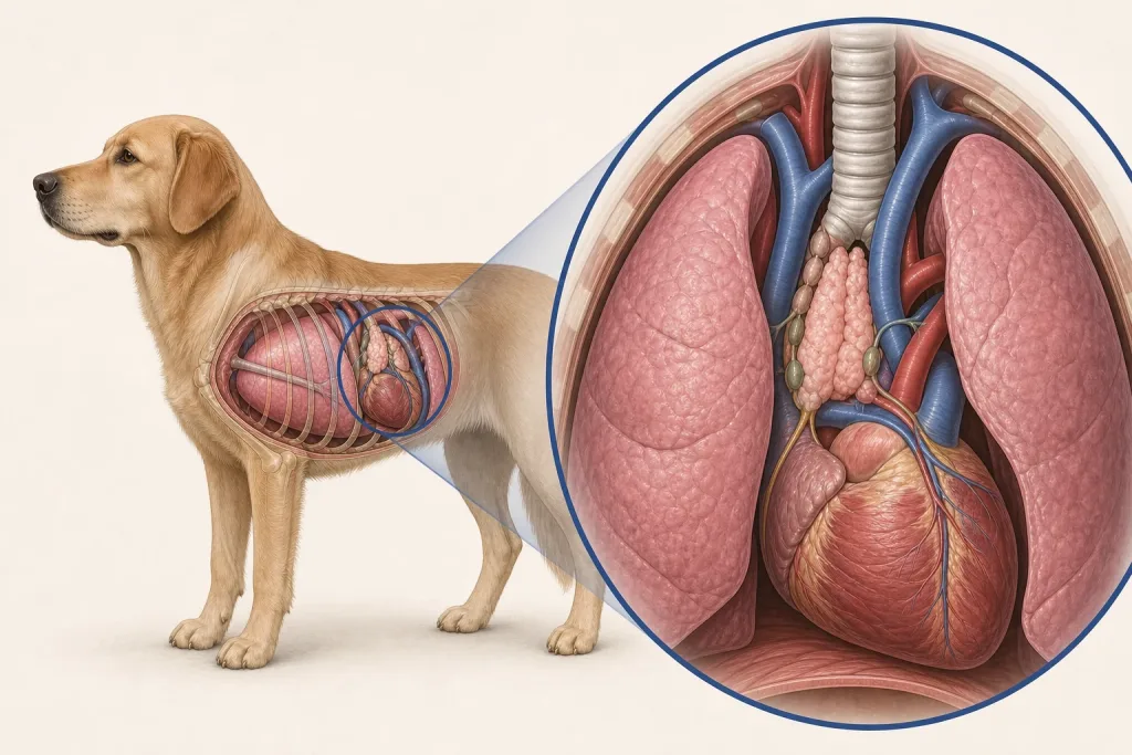
Overview
Mediastinal Lymphoma is lymphoid cancer centered in lymphatic or thymic tissue in the front portion of the chest. It can compress airways and major vessels or cause chest fluid, and a cranial mediastinal mass also has differentials such as thymoma and other tumors.
Common signs
Owners may notice rapid or difficult breathing, cough, exercise intolerance, regurgitation, swelling of the head or neck, weakness, increased thirst or urination from high calcium, or reduced appetite. None of these signs is specific to mediastinal lymphoma, so a veterinary examination and appropriate testing are needed before naming the problem.
Common locations
The cranial mediastinum contains the thymus, lymph nodes, vessels, trachea, and esophagus; lymphoma may also involve marrow, blood, spleen, liver, or other nodes. Anatomy affects what can be sampled safely, whether a complete first surgery is realistic, and which nearby function needs protection.
Diagnosis discussion
Chest imaging, careful needle sampling when safe, flow cytometry or immunophenotyping, bloodwork including calcium, and additional staging may be recommended. Stabilization takes priority when breathing is compromised. The diagnostic plan should answer a decision: whether tissue type, local extent, stage, or patient health will change the next option. An imaging impression is not the same as a histologic diagnosis.
Urgency and when to contact a veterinarian
Arrange a prompt veterinary appointment for persistent or progressive signs. Seek emergency veterinary care for any of these signs: labored breathing, blue or gray gums, collapse, inability to swallow, rapidly increasing head or neck swelling, or profound weakness. Call ahead when possible so the receiving team can prepare; do not delay emergency transport to take photographs or complete a log.
Treatment discussion
Systemic therapy is commonly discussed after classification. Fluid drainage, oxygen, management of hypercalcemia, and other supportive care may be required before or during treatment; radiation or other options are case dependent. The team should explain the goal of each option—diagnosis, local control, systemic control, symptom relief, or emergency stabilization—along with likely burdens, costs, travel, rechecks, and alternatives.
Home monitoring
Keep one dated home record of sleeping respiratory rate, effort, cough, neck or facial swelling, swallowing, thirst, urination, appetite, energy, temperature if directed, and treatment side effects. Use the same measurement or observation method each time and share trends with the veterinary team. Home tracking supports planned rechecks; it cannot determine grade, margins, or metastasis.
Questions to ask your veterinarian
- Does imaging favor lymphoma, thymoma, or another mediastinal process?
- Can a sample be obtained without worsening breathing or bleeding risk?
- Which immunophenotyping test will guide the treatment discussion?
- Is high calcium or chest fluid contributing to the current urgency?