Canine Glioma: MRI Defines the Lesion, but Tissue Defines the Tumor
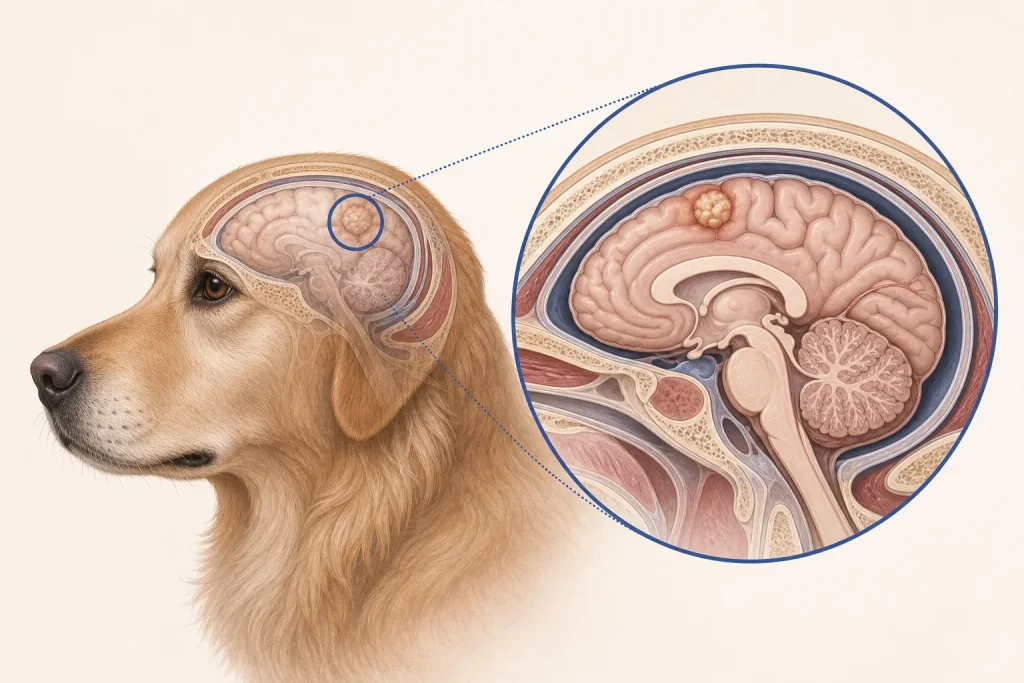
Overview
Glioma is a group of primary brain tumors arising from glial cells, including astrocytic and oligodendroglial tumors. Gliomas often grow within brain tissue, and imaging can strongly suggest a glioma but cannot reliably provide every grade or subtype required for prognosis or trial eligibility.
Common signs
Owners may notice seizures, altered behavior, circling, weakness on one side, vision change, balance problems, head pressing, or a progressive change in learned behavior. None of these signs is specific to glioma, so a veterinary examination and appropriate testing are needed before naming the problem.
Common locations
They commonly affect the cerebral hemispheres but can occur elsewhere in the central nervous system; the exact lobe and relationship to critical pathways shape signs and options. Anatomy affects what can be sampled safely, whether a complete first surgery is realistic, and which nearby function needs protection.
Diagnosis discussion
Neurologic localization, MRI with contrast, laboratory evaluation, and specialist discussion of stereotactic biopsy or other tissue acquisition may be appropriate. Other inflammatory, vascular, and neoplastic lesions can mimic a glioma. The diagnostic plan should answer a decision: whether tissue type, local extent, stage, or patient health will change the next option. An imaging impression is not the same as a histologic diagnosis.
Urgency and when to contact a veterinarian
Arrange a prompt veterinary appointment for persistent or progressive signs. Seek emergency veterinary care for any of these signs: prolonged or clustered seizures, inability to recover, sudden collapse, severe breathing change, inability to stand, or rapidly worsening mentation. Call ahead when possible so the receiving team can prepare; do not delay emergency transport to take photographs or complete a log.
Treatment discussion
Radiation, surgery or minimally invasive approaches in selected centers, clinical trials, seizure and swelling control, and palliative care may be considered. Claims about experimental therapies should be separated from established care and trial consent. The team should explain the goal of each option—diagnosis, local control, systemic control, symptom relief, or emergency stabilization—along with likely burdens, costs, travel, rechecks, and alternatives.
Home monitoring
Keep one dated home record of seizure pattern, recovery, walking, vision, behavior, sleep, appetite, medication effects, and whether daily function is stable between episodes. Use the same measurement or observation method each time and share trends with the veterinary team. Home tracking supports planned rechecks; it cannot determine grade, margins, or metastasis.
Questions to ask your veterinarian
- Which inflammatory or vascular diseases remain in the MRI differential?
- Would biopsy change treatment selection or clinical-trial eligibility?
- What are the goals and burdens of radiation or other local therapy?
- What seizure duration or cluster pattern requires emergency medication or transport?