Canine Choroid Plexus Tumors: Fluid Flow and Tumor Type Both Matter
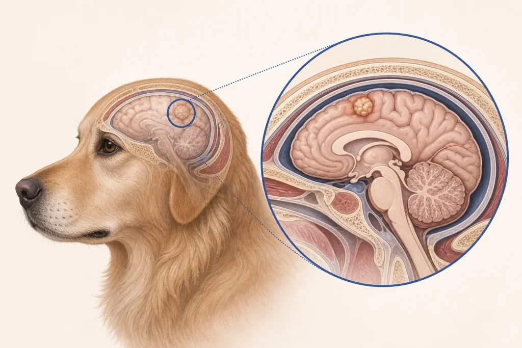
Overview
Choroid Plexus Tumors are papillary tumors arising from tissue that produces cerebrospinal fluid inside the brain’s ventricles. They may obstruct fluid flow, cause hydrocephalus, invade nearby tissue, or spread through cerebrospinal fluid pathways; imaging location is informative but does not guarantee subtype.
Common signs
Owners may notice seizures, altered alertness, circling, imbalance, neck pain, vision change, weakness, or intermittent episodes associated with pressure inside the skull. None of these signs is specific to choroid plexus tumors, so a veterinary examination and appropriate testing are needed before naming the problem.
Common locations
The lateral, third, and fourth ventricles are possible sites, and tumor position determines whether the forebrain, brainstem, or cerebellar signs dominate. Anatomy affects what can be sampled safely, whether a complete first surgery is realistic, and which nearby function needs protection.
Diagnosis discussion
Neurologic examination and MRI are central. The team may discuss CSF flow, hydrocephalus, staging of the neuraxis, and whether surgery or biopsy can safely provide histopathology. The diagnostic plan should answer a decision: whether tissue type, local extent, stage, or patient health will change the next option. An imaging impression is not the same as a histologic diagnosis.
Urgency and when to contact a veterinarian
Arrange a prompt veterinary appointment for persistent or progressive signs. Seek emergency veterinary care for any of these signs: repeated vomiting with neurologic change, prolonged seizures, sudden inability to stand, severe obtundation, breathing change, or rapid deterioration. Call ahead when possible so the receiving team can prepare; do not delay emergency transport to take photographs or complete a log.
Treatment discussion
Surgery, radiation, procedures to manage obstructed fluid flow, symptom-control medication, and palliative care may be considered. Planning should distinguish temporary pressure relief from tumor-directed treatment. The team should explain the goal of each option—diagnosis, local control, systemic control, symptom relief, or emergency stabilization—along with likely burdens, costs, travel, rechecks, and alternatives.
Home monitoring
Keep one dated home record of seizures, gait and balance, head or neck comfort, alertness, sleep, vision, appetite, medication effects, and recovery after any pressure-related episode. Use the same measurement or observation method each time and share trends with the veterinary team. Home tracking supports planned rechecks; it cannot determine grade, margins, or metastasis.
Questions to ask your veterinarian
- Is cerebrospinal fluid flow obstructed or hydrocephalus present?
- Should the full neuraxis be imaged for dissemination?
- Can surgery provide both decompression and representative tissue?
- Which changes indicate rising intracranial pressure needs urgent care?