Canine Spinal Nephroblastoma: A Young Dog With Progressive Hindlimb Signs
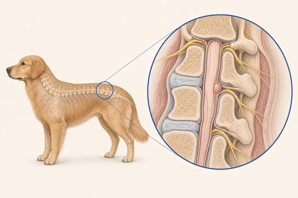
Overview
Spinal Nephroblastoma is a rare spinal tumor reported most often in young dogs and thought to arise from embryonic tissue remnants. It commonly develops near the thoracolumbar cord, but disc disease, congenital disorders, inflammation, trauma, and other tumors can produce similar neurologic signs.
Common signs
Owners may notice progressive hindlimb weakness, incoordination, nail scuffing, difficulty rising, spinal pain, muscle loss, altered tail function, or urinary and fecal changes. None of these signs is specific to spinal nephroblastoma, so a veterinary examination and appropriate testing are needed before naming the problem.
Common locations
The thoracolumbar spinal canal is a characteristic location, often outside the cord but within its coverings, where compression may progress despite a young age. Anatomy affects what can be sampled safely, whether a complete first surgery is realistic, and which nearby function needs protection.
Diagnosis discussion
Neurologic localization and MRI are central, followed by specialist assessment for surgical exploration and histopathology. Age and location may raise suspicion, but neither is a definitive diagnosis. The diagnostic plan should answer a decision: whether tissue type, local extent, stage, or patient health will change the next option. An imaging impression is not the same as a histologic diagnosis.
Urgency and when to contact a veterinarian
Arrange a prompt veterinary appointment for persistent or progressive signs. Seek emergency veterinary care for any of these signs: rapid loss of walking, inability to urinate, severe pain, breathing weakness, or sudden neurologic decline. Call ahead when possible so the receiving team can prepare; do not delay emergency transport to take photographs or complete a log.
Treatment discussion
Surgical decompression and removal may be discussed when feasible, sometimes with radiation or other adjunctive care depending on pathology and residual disease. Rehabilitation and bladder support need an explicit plan. The team should explain the goal of each option—diagnosis, local control, systemic control, symptom relief, or emergency stabilization—along with likely burdens, costs, travel, rechecks, and alternatives.
Home monitoring
Keep one dated home record of hindlimb strength, falls, paw placement, nail wear, pain, ability to rise, urination, stool control, sleep, and changes captured in brief dated videos. Use the same measurement or observation method each time and share trends with the veterinary team. Home tracking supports planned rechecks; it cannot determine grade, margins, or metastasis.
Questions to ask your veterinarian
- Does the age and thoracolumbar location support this diagnosis, and what alternatives remain?
- Can surgery safely decompress the cord and obtain representative tissue?
- Would postoperative radiation be discussed if residual disease remains?
- What bladder and mobility support is needed before and after referral?