Canine Synovial Sarcoma: Persistent Joint-Area Swelling Needs Tissue Diagnosis
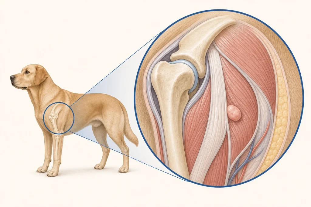
Overview
Synovial Sarcoma is a malignant mesenchymal tumor that develops near joints and may involve surrounding soft tissue or bone. The historical name does not mean every lesion arises from true joint lining, and chronic orthopedic or inflammatory disease can mimic it.
Common signs
Owners may notice progressive lameness, firm swelling near a joint, pain, reduced range of motion, muscle loss, reluctance to exercise, or a mass that persists despite routine orthopedic treatment. None of these signs is specific to synovial sarcoma, so a veterinary examination and appropriate testing are needed before naming the problem.
Common locations
Large limb joints are typical regions, but exact origin from periarticular soft tissue, tendon sheath, joint capsule, or bone must be mapped. Anatomy affects what can be sampled safely, whether a complete first surgery is realistic, and which nearby function needs protection.
Diagnosis discussion
Orthopedic examination, radiographs, CT or MRI, carefully planned core or incisional biopsy, histopathology with ancillary testing, and staging may be recommended before definitive surgery. The diagnostic plan should answer a decision: whether tissue type, local extent, stage, or patient health will change the next option. An imaging impression is not the same as a histologic diagnosis.
Urgency and when to contact a veterinarian
Arrange a prompt veterinary appointment for persistent or progressive signs. Seek urgent veterinary care for any of these signs: sudden non-weight-bearing pain, suspected fracture, uncontrolled bleeding from an ulcerated mass, severe swelling, or rapid decline. Call ahead when possible so the receiving team can prepare; do not delay emergency transport to take photographs or complete a log.
Treatment discussion
Limb-sparing wide removal or amputation may be discussed according to location and margins. Radiation, systemic therapy, pain management, and rehabilitation are considered case by case; an unplanned shell-out can limit later options. The team should explain the goal of each option—diagnosis, local control, systemic control, symptom relief, or emergency stabilization—along with likely burdens, costs, travel, rechecks, and alternatives.
Home monitoring
Keep one dated home record of lameness score, joint use, swelling dimensions, pain at rest, sleep, ability to rise, muscle symmetry, appetite, and medication effects. Use the same measurement or observation method each time and share trends with the veterinary team. Home tracking supports planned rechecks; it cannot determine grade, margins, or metastasis.
Questions to ask your veterinarian
- Is the lesion centered in bone, the joint, or periarticular soft tissue?
- Where should the biopsy tract lie so it can be removed with definitive surgery?
- Can limb-sparing margins preserve useful function, or is amputation more predictable?
- Which staging and pathology findings would change systemic therapy discussion?