Canine Urethral Carcinoma: Straining With Little Urine Can Become an Emergency
Overview
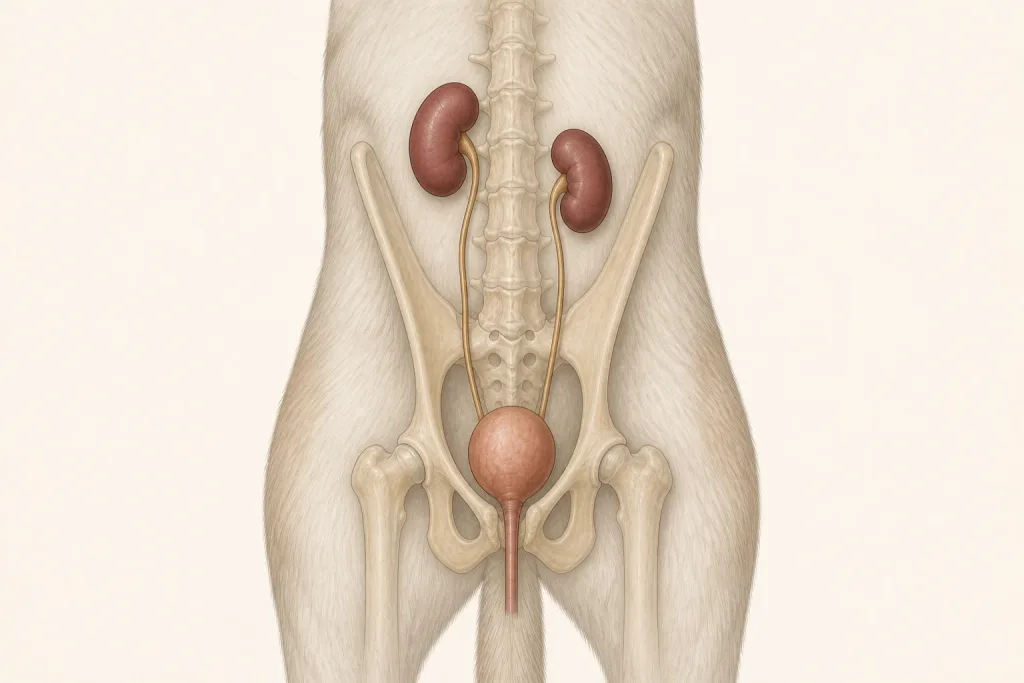
Urethral Carcinoma is a malignant epithelial tumor arising in the tube that carries urine out of the bladder. It can obstruct urine flow and may be part of urothelial carcinoma involving the bladder neck, urethra, or prostate; infection and stones can cause similar signs.
Common signs
Owners may notice frequent urination attempts, straining, blood in urine, a weak or interrupted stream, accidents, pain, recurrent urinary infection, licking, or progressive inability to pass urine. None of these signs is specific to urethral carcinoma, so a veterinary examination and appropriate testing are needed before naming the problem.
Common locations
The proximal or distal urethra may be affected, with sex-specific anatomy influencing examination, cystoscopy, biopsy, stenting, and surgical options. Anatomy affects what can be sampled safely, whether a complete first surgery is realistic, and which nearby function needs protection.
Diagnosis discussion
Urinalysis and culture, ultrasound, contrast imaging or CT, cystoscopy, carefully selected sampling, and staging may be proposed. The team avoids procedures that could seed tumor when a safer validated test is available. The diagnostic plan should answer a decision: whether tissue type, local extent, stage, or patient health will change the next option. An imaging impression is not the same as a histologic diagnosis.
Urgency and when to contact a veterinarian
Arrange a prompt veterinary appointment for persistent or progressive signs. Seek emergency veterinary care for any of these signs: repeated straining with no urine, a painful distended abdomen, vomiting, collapse, profound weakness, or rapidly worsening pain. Call ahead when possible so the receiving team can prepare; do not delay emergency transport to take photographs or complete a log.
Treatment discussion
Medical oncology, radiation, surgery in selected sites, stenting or diversion procedures, and palliative care may be discussed. Restoring urine flow and controlling infection and pain can be more urgent than completing every staging test. The team should explain the goal of each option—diagnosis, local control, systemic control, symptom relief, or emergency stabilization—along with likely burdens, costs, travel, rechecks, and alternatives.
Home monitoring
Keep one dated home record of number of attempts, stream strength and duration, urine volume and color, pain, accidents, appetite, water intake, and whether urine can be passed at every outing. Use the same measurement or observation method each time and share trends with the veterinary team. Home tracking supports planned rechecks; it cannot determine grade, margins, or metastasis.
Questions to ask your veterinarian
- Is urine flow currently adequate, or is decompression needed first?
- Where does the lesion begin and end on imaging or cystoscopy?
- Which sampling approach minimizes seeding and obstruction risk?
- Would a stent, catheter, surgery, radiation, or medication best address current goals?