Canine Vaginal and Vulvar Tumors: Protruding Tissue Is Not a Visual Diagnosis
Overview
Vaginal and Vulvar Tumors are a group of smooth-muscle, epithelial, transmissible, and other tumors affecting the vagina, vestibule, or vulva. Benign leiomyoma is common in this region, but malignant tumors, vaginal hyperplasia, prolapse, inflammation, and trauma can appear similar.
Common signs
Owners may notice a protruding mass, bleeding or discharge, licking, straining to urinate or defecate, pain, recurrent urinary infection, mating difficulty, or a lump noticed during examination. None of these signs is specific to vaginal and vulvar tumors, so a veterinary examination and appropriate testing are needed before naming the problem.
Common locations
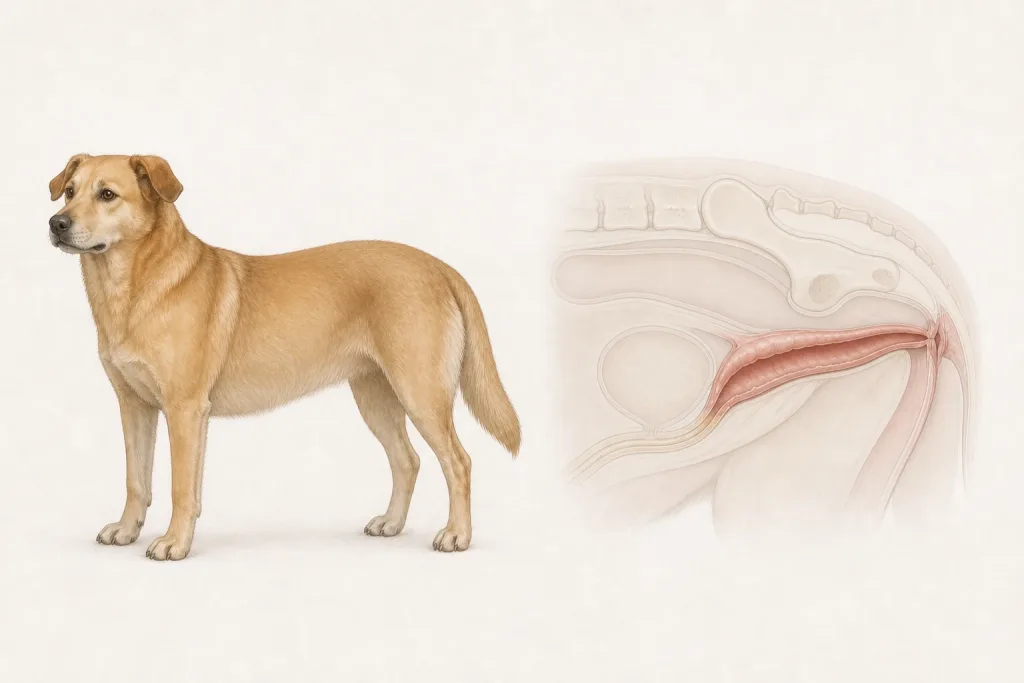
The vulva, vestibule, and deeper vagina have different relationships to the urethra, rectum, cervix, and pelvic canal, so examination under sedation and imaging may be needed. Anatomy affects what can be sampled safely, whether a complete first surgery is realistic, and which nearby function needs protection.
Diagnosis discussion
Reproductive history, rectal and vaginal examination, cytology or biopsy, imaging for deep extension, and staging based on histology may be discussed. Contagious transmissible venereal tumor remains a specific differential in relevant travel and contact histories. The diagnostic plan should answer a decision: whether tissue type, local extent, stage, or patient health will change the next option. An imaging impression is not the same as a histologic diagnosis.
Urgency and when to contact a veterinarian
Arrange a prompt veterinary appointment for persistent or progressive signs. Seek urgent veterinary care for any of these signs: inability to urinate, heavy bleeding, dark or severely swollen protruding tissue, collapse, severe pain, or rapid enlargement. Call ahead when possible so the receiving team can prepare; do not delay emergency transport to take photographs or complete a log.
Treatment discussion
Surgical removal, ovariohysterectomy for hormone-responsive disease, systemic therapy for transmissible or other chemosensitive tumors, radiation, or comfort care may be considered after classification. The team should explain the goal of each option—diagnosis, local control, systemic control, symptom relief, or emergency stabilization—along with likely burdens, costs, travel, rechecks, and alternatives.
Home monitoring
Keep one dated home record of bleeding or discharge, tissue color and trauma, urine stream, straining, stool passage, licking, pain, appetite, and mass recurrence. Use the same measurement or observation method each time and share trends with the veterinary team. Home tracking supports planned rechecks; it cannot determine grade, margins, or metastasis.
Questions to ask your veterinarian
- Where is the attachment relative to the urethra and rectum?
- Could prolapse, hyperplasia, or transmissible venereal tumor mimic this lesion?
- Will biopsy or removal preserve continence and later treatment options?
- Which bleeding or urinary changes require same-day care?