Canine Sertoli Cell Tumor: A Testicular Mass Can Produce Whole-Body Hormonal Signs
Overview
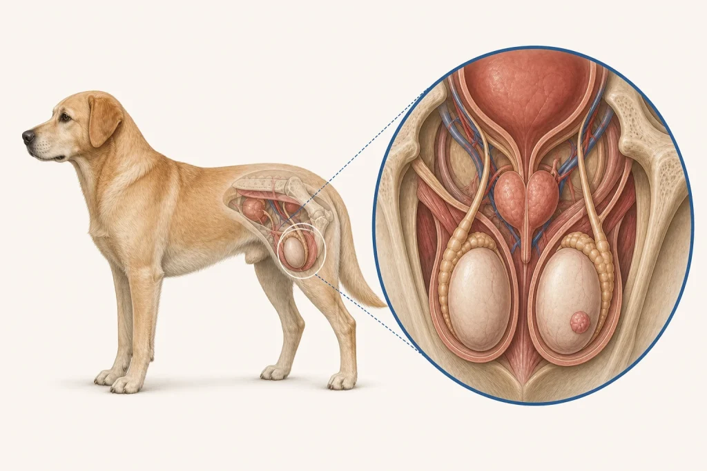
Sertoli Cell Tumor is a sex-cord stromal tumor arising from Sertoli cells within a testicle. Many remain local, but some spread and some release estrogen, particularly when a testicle is retained in the abdomen or groin; hormone effects can suppress bone marrow.
Common signs
Owners may notice testicular enlargement or asymmetry, a groin or abdominal mass, symmetric hair loss, darkened skin, enlarged nipples, attraction of male dogs, a pendulous prepuce, weakness, bruising, fever, or pale gums. None of these signs is specific to sertoli cell tumor, so a veterinary examination and appropriate testing are needed before naming the problem.
Common locations
A scrotal, inguinal, or abdominal retained testicle may be affected; the opposite testicle, prostate, regional nodes, and marrow effects can require evaluation. Anatomy affects what can be sampled safely, whether a complete first surgery is realistic, and which nearby function needs protection.
Diagnosis discussion
Physical examination, blood counts, abdominal or scrotal ultrasound, staging as indicated, and histopathology after removal are discussed. Cytopenias require careful perioperative planning and may persist after the hormone source is removed. The diagnostic plan should answer a decision: whether tissue type, local extent, stage, or patient health will change the next option. An imaging impression is not the same as a histologic diagnosis.
Urgency and when to contact a veterinarian
Arrange a prompt veterinary appointment for persistent or progressive signs. Seek urgent veterinary care for any of these signs: very pale gums, uncontrolled bleeding, fever with severe weakness, collapse, breathing difficulty, or sudden painful testicular swelling. Call ahead when possible so the receiving team can prepare; do not delay emergency transport to take photographs or complete a log.
Treatment discussion
Removal of both testicles is commonly recommended, with oncology and supportive treatment guided by pathology, spread, and severity of estrogen-related marrow suppression. The team should explain the goal of each option—diagnosis, local control, systemic control, symptom relief, or emergency stabilization—along with likely burdens, costs, travel, rechecks, and alternatives.
Home monitoring
Keep one dated home record of gum color, bruising, fever if instructed, energy, appetite, hair and skin change, testicular or groin size, urination, and laboratory recheck dates. Use the same measurement or observation method each time and share trends with the veterinary team. Home tracking supports planned rechecks; it cannot determine grade, margins, or metastasis.
Questions to ask your veterinarian
- Is a retained abdominal or inguinal testicle present?
- Do blood counts show estrogen-related marrow suppression?
- Should both testicles and any suspicious nodes be removed or sampled?
- What blood-count trend is expected after surgery, and when is transfusion support considered?