Canine Acute Leukemia: Immature Blood Cells Can Crowd Out Normal Marrow
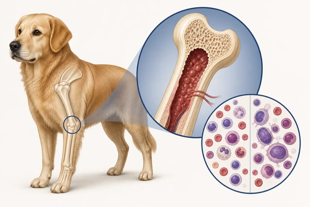
Overview
Acute Leukemia is a rapidly progressive cancer of immature blood-forming cells, classified by lineage and immunophenotype. Abnormal blast cells can replace normal marrow production, leading to anemia, infection risk, and bleeding; a high or low white-cell count alone does not establish the diagnosis.
Common signs
Owners may notice marked lethargy, pale gums, fever, bruising or bleeding, reduced appetite, weight loss, enlarged nodes or organs, recurrent infection, breathing effort, or sudden weakness. None of these signs is specific to acute leukemia, so a veterinary examination and appropriate testing are needed before naming the problem.
Common locations
Bone marrow and blood are central, with possible involvement of spleen, liver, lymph nodes, skin, nervous system, or other organs depending on lineage. Anatomy affects what can be sampled safely, whether a complete first surgery is realistic, and which nearby function needs protection.
Diagnosis discussion
Repeat CBC with smear review, chemistry, coagulation testing, infectious-disease evaluation when relevant, bone-marrow aspiration or biopsy, flow cytometry, cytochemistry, and molecular tests may be needed to identify lineage. The diagnostic plan should answer a decision: whether tissue type, local extent, stage, or patient health will change the next option. An imaging impression is not the same as a histologic diagnosis.
Urgency and when to contact a veterinarian
Arrange a prompt veterinary appointment for persistent or progressive signs. Seek emergency veterinary care for any of these signs: uncontrolled bleeding, fever with profound weakness, collapse, breathing difficulty, very pale gums, seizures, or inability to stand. Call ahead when possible so the receiving team can prepare; do not delay emergency transport to take photographs or complete a log.
Treatment discussion
Urgent specialist-directed systemic therapy and intensive supportive care may be discussed. Transfusion, antimicrobial, nausea, nutrition, and bleeding support are individualized; prognosis and treatment burden should be communicated plainly. The team should explain the goal of each option—diagnosis, local control, systemic control, symptom relief, or emergency stabilization—along with likely burdens, costs, travel, rechecks, and alternatives.
Home monitoring
Keep one dated home record of gum color, bruising, bleeding, temperature if directed, appetite, breathing, energy, infection exposure, medication effects, and exact laboratory recheck dates. Use the same measurement or observation method each time and share trends with the veterinary team. Home tracking supports planned rechecks; it cannot determine grade, margins, or metastasis.
Questions to ask your veterinarian
- Which cell lineage is supported by flow cytometry or other classification tests?
- Could infection, immune disease, or treatment effect explain the blood changes?
- What immediate transfusion or infection support is needed?
- What findings would pause or change systemic therapy?