Canine Mammary Tumors: A Lump Along the Mammary Chain Needs Pathology
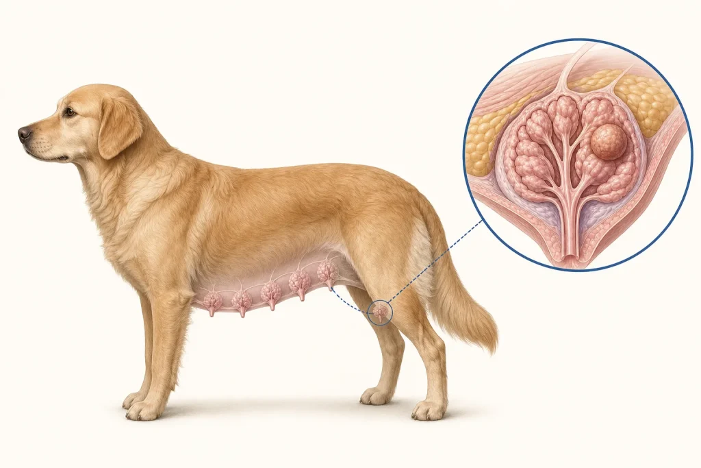
Overview
Canine mammary tumors include many benign and malignant subtypes. Clinical appearance does not reliably distinguish them; histopathology after biopsy or removal defines the diagnosis and important prognostic features.
Common signs
One or more nodules along the mammary chain, change in nipple or gland shape, discharge, ulceration, bleeding, pain or swelling. Advanced disease may be accompanied by reduced appetite, weight loss or breathing signs.
Common locations
Any mammary gland from the chest to the groin. Multiple glands can be affected, and regional lymphatic drainage differs by location.
Diagnosis discussion
The examination should map every mammary mass and regional lymph node. Cytology may help rule out some differentials but often cannot fully classify mammary tumors. Histopathology is central. Chest imaging, bloodwork and selective abdominal or nodal evaluation may be recommended before treatment.
Urgency and when to contact a veterinarian
Book a prompt assessment for a mammary lump. Rapid diffuse swelling, heat, severe pain, ulceration, uncontrolled bleeding, breathing difficulty or marked illness needs urgent evaluation.
Treatment discussion
Surgical extent depends on species, number, size and location of masses and patient health. Every removed mass should be identified and submitted for histopathology. Additional systemic treatment is case-specific. Inflammatory mammary carcinoma is managed differently and is generally not treated with routine surgery.
Home monitoring
Once monthly, gently inspect the full mammary chain without squeezing masses. Record size and skin change. After surgery, use the prescribed collar, restrict activity, inspect the incision and attend pathology and surveillance visits.
Questions to ask your veterinarian
- How many distinct masses are present, and will each be labeled for pathology?
- What surgical extent is appropriate for their size, location and number?
- Is there any concern for inflammatory mammary carcinoma?
- What do tumor type, grade, margins and stage mean for follow-up?