Canine Mast Cell Tumor: From a Changing Lump to a Tissue Diagnosis
Overview
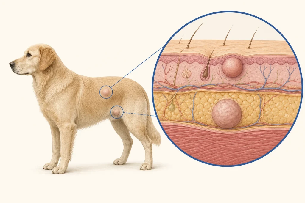
Mast cell tumors arise from immune cells that contain histamine and other inflammatory mediators. They most often develop in or under the skin and range from low-risk, locally controlled tumors to aggressive disease, so appearance alone cannot predict behavior.
Common signs
A new or changing skin lump; waxing and waning size; redness, bruising, itching, licking, ulceration or bleeding. Some dogs develop vomiting, poor appetite or dark stool when inflammatory mediators affect the gastrointestinal tract.
Common locations
Skin and subcutaneous tissue anywhere on the body, including the trunk, limbs, head and neck. Internal mast cell disease is a different and generally more serious presentation.
Diagnosis discussion
Fine-needle aspiration and cytology often identify a mast cell tumor. Histopathology is needed for grade and margin assessment. Depending on the pathology, site and clinical findings, the veterinarian may recommend regional lymph-node sampling, bloodwork and selective imaging.
Urgency and when to contact a veterinarian
Book a prompt appointment for every new or changing mass. Use urgent or emergency care for collapse, breathing difficulty, uncontrolled bleeding, repeated vomiting, black/tarry stool, marked weakness or rapidly increasing swelling.
Treatment discussion
Planned surgery is commonly used for local control. Radiation may be considered for selected incompletely removed or difficult tumors. Chemotherapy or targeted therapy may be discussed for high-risk, recurrent, nonresectable or metastatic disease. The plan depends on grade, stage, location, margins and patient factors.
Home monitoring
Do not squeeze the mass. Photograph it beside a ruler on a consistent schedule and record redness, surface change, licking, appetite, vomiting, stool color, energy and prescribed medication effects. Follow the clinic's recheck plan even if the site looks quiet.
Questions to ask your veterinarian
- Can this mass be sampled today, and is cytology likely to be sufficient?
- If surgery is considered, what margins and reconstruction plan are appropriate before the first operation?
- Should the draining lymph node be sampled even if it is not enlarged?
- Which pathology findings would change staging or additional treatment?