Canine Thymoma: An Anterior Chest Mass With Neuromuscular Clues
Overview
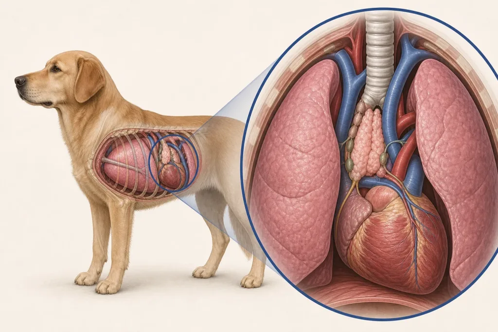
A thymoma arises from thymic epithelial tissue in the front portion of the chest. Lymphoma and other mediastinal masses can look similar on imaging, and some thymomas are associated with paraneoplastic myasthenia gravis.
Common signs
Cough, rapid or difficult breathing, exercise intolerance, regurgitation, swallowing difficulty, generalized weakness, or an enlarged chest silhouette may occur. Some dogs have a mass found before obvious signs.
Common locations
The thymus lies in the cranial mediastinum. Tumor contact with the heart, major vessels, airway, lungs, and nerves affects surgical and anesthesia planning.
Diagnosis discussion
Chest radiographs and CT help define the mass; cytology or biopsy may distinguish thymoma from lymphoma or other tumors. Dogs with regurgitation or weakness may need testing for myasthenia gravis and evaluation of esophageal dilation before anesthesia.
Urgency and when to contact a veterinarian
Labored breathing, blue or pale gums, collapse, repeated regurgitation with cough or fever, or sudden inability to stand needs urgent care because respiratory compromise or aspiration pneumonia is possible.
Treatment discussion
Surgical removal may be considered for a resectable thymoma. Radiation or palliative treatment can be discussed for nonresectable disease. Associated myasthenia gravis, megaesophagus, aspiration risk, and nutrition may require separate ongoing management.
Home monitoring
Track resting breathing, cough, regurgitation versus vomiting, swallowing, appetite, weight, weakness, and temperature if directed. Feed positioning or texture changes should follow the veterinary team’s plan.
Questions to ask your veterinarian
- How confident are we that this is thymoma rather than lymphoma?
- Does CT show invasion that changes surgical feasibility?
- Should the dog be tested for myasthenia gravis or megaesophagus?
- What aspiration-pneumonia signs require immediate care?