Canine Meningioma: Location, Imaging, and Neurologic Function Guide Decisions
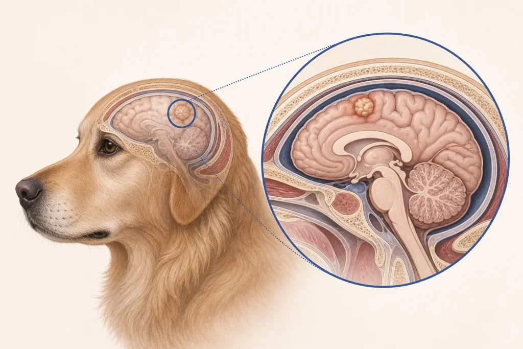
Overview
Meningioma is a tumor arising from the membranes surrounding the brain or spinal cord. Many canine meningiomas grow as extra-axial masses, but their effect can be serious because pressure and invasion occur within a fixed skull; MRI appearance is suggestive, not histologic proof.
Common signs
Owners may notice new seizures, circling, behavior change, vision loss, head pressing, imbalance, weakness on one side, neck or back pain, or progressive difficulty walking. None of these signs is specific to meningioma, so a veterinary examination and appropriate testing are needed before naming the problem.
Common locations
Meningiomas can develop over the cerebral hemispheres, near the skull base, around the brainstem, or along the spinal meninges; location drives signs and accessibility. Anatomy affects what can be sampled safely, whether a complete first surgery is realistic, and which nearby function needs protection.
Diagnosis discussion
A neurologic examination, bloodwork to evaluate seizure mimics and anesthesia fitness, MRI, sometimes CSF analysis, and biopsy or surgical histopathology in selected cases may be discussed. The diagnostic plan should answer a decision: whether tissue type, local extent, stage, or patient health will change the next option. An imaging impression is not the same as a histologic diagnosis.
Urgency and when to contact a veterinarian
Arrange a prompt veterinary appointment for persistent or progressive signs. Seek emergency veterinary care for any of these signs: a seizure lasting several minutes, repeated seizures without full recovery, sudden inability to stand, severe disorientation, breathing difficulty, or rapid neurologic decline. Call ahead when possible so the receiving team can prepare; do not delay emergency transport to take photographs or complete a log.
Treatment discussion
Surgery, radiation, symptom-control medication, or palliative care may be considered according to location, imaging, tissue diagnosis, age, comorbidities, and caregiver goals. Medication may control swelling or seizures without removing the mass. The team should explain the goal of each option—diagnosis, local control, systemic control, symptom relief, or emergency stabilization—along with likely burdens, costs, travel, rechecks, and alternatives.
Home monitoring
Keep one dated home record of seizure date and duration, recovery time, gait videos, circling, vision, sleep, appetite, medication timing, sedation, and ability to perform normal activities safely. Use the same measurement or observation method each time and share trends with the veterinary team. Home tracking supports planned rechecks; it cannot determine grade, margins, or metastasis.
Questions to ask your veterinarian
- Which MRI features support meningioma, and what alternatives remain?
- Is the location surgically accessible without unacceptable functional risk?
- Would tissue confirmation change radiation or medication choices?
- What written seizure action plan should we follow after hours?