Anal Sac Adenocarcinoma: Connecting Rectal Examination, Calcium, and Abdominal Nodes
Summary
A focused diagnostic and staging pathway for a tumor whose local size, calcium effects, and nodal spread may tell different parts of the story.
Article
Educational medical disclaimer: This material is general education and cannot diagnose an anal sac tumor, interpret calcium results, or recommend surgery for a particular dog. Straining without producing stool or urine, repeated vomiting, profound weakness, collapse, or rapid decline needs urgent veterinary attention.
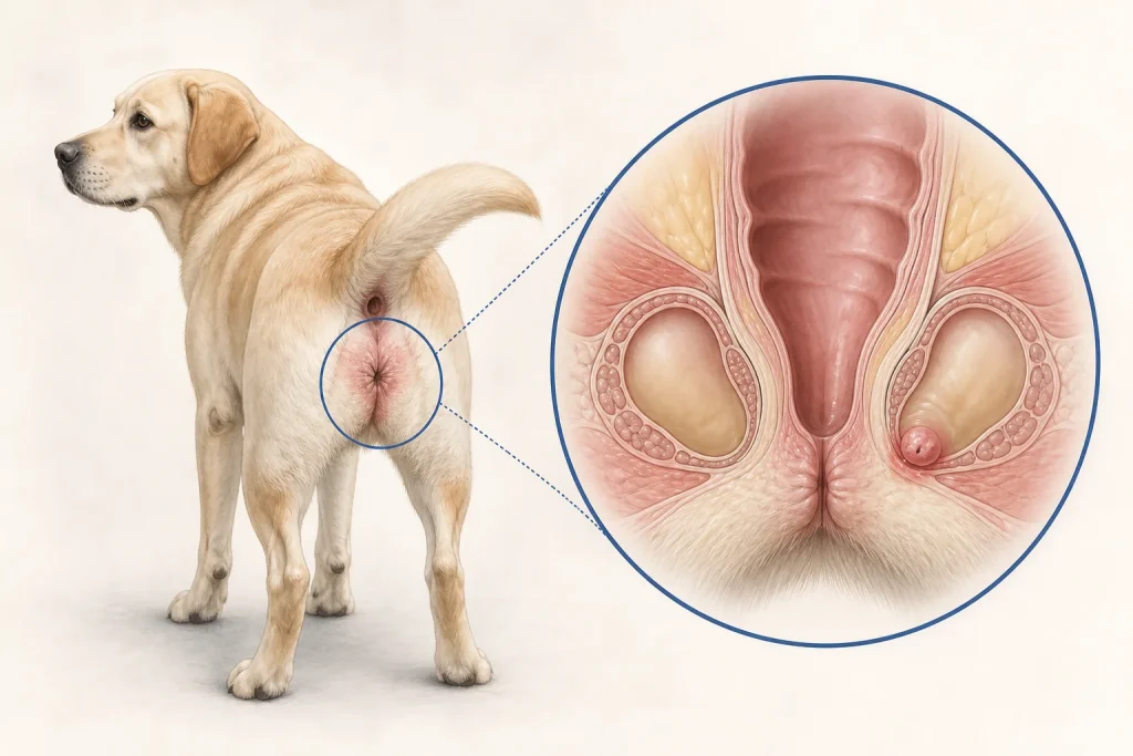
Apocrine gland anal sac adenocarcinoma can be discovered because a dog scoots or strains, during evaluation of increased thirst, or on a routine rectal examination. Some tumors are small at the primary site while abdominal lymph nodes are more conspicuous. Some dogs develop clinically important high calcium, while others do not. A coherent workup therefore connects three tracks: local anatomy, tumor identity, and whole-dog effects.
The rectal examination still matters
The anal sacs sit beside the anus, and a mass may not be visible from the outside. Digital rectal examination can identify asymmetry or firmness and may also reveal changes that affect defecation. The veterinarian will assess the skin, perineum, regional structures, and accessible lymph nodes, then evaluate general hydration, strength, heart rate, and other findings. Scooting alone is common and nonspecific; it should not be treated as proof of cancer.
Obtain cells or tissue
A fine-needle sample of a primary mass or accessible node can often help establish a diagnosis. Histopathology provides tissue architecture and may follow biopsy or surgery. The sampling plan depends on size, location, bleeding risk, whether infection is present, and how the result will change care. If cytology is inconclusive, repeating a better-targeted sample or obtaining tissue can be more informative than simply labeling the mass benign.
Measure calcium carefully
Some anal sac tumors are associated with parathyroid hormone-related protein and hypercalcemia. High calcium can contribute to increased drinking and urination, appetite loss, constipation, vomiting, weakness, and kidney injury, but those signs have many other causes. Total serum calcium can be affected by proteins and other factors. A study evaluating dogs with total hypercalcemia or total hypocalcemia found that total-calcium-based prediction did not consistently reflect directly measured ionized calcium. Clinicians may therefore confirm an unexpected result with ionized calcium and interpret it with kidney values, hydration, medications, and the broader differential.
Hypercalcemia does not identify the tumor by itself. Other malignancies and noncancerous disorders can cause it. Conversely, a normal calcium value does not exclude an anal sac tumor. The laboratory result is one part of the map, not a shortcut around examination and tissue diagnosis.
Stage the relevant anatomy
The American College of Veterinary Surgeons describes abdominal imaging as part of evaluation because sublumbar lymph nodes can be involved. Thoracic imaging and assessment of other sites may be recommended according to findings and goals. Enlarged nodes can have noncancerous causes, and normal-sized nodes cannot be assumed negative. Image-guided sampling may be considered when nodal status would alter surgery, radiation, systemic therapy, or prognosis.
Discuss treatment as a set of jobs
Surgery may target the primary mass and, in selected cases, affected lymph nodes. Radiation or systemic therapy may be discussed depending on distribution, pathology, symptoms, and specialist judgment. Supportive care can address pain, nausea, constipation, hydration, kidney risk, or other burdens. Each option has potential complications and practical demands. No article can decide whether the expected benefit outweighs those burdens for an individual dog.
Questions for the care conference
- Was the diagnosis confirmed from the primary site, a node, or both?
- Was ionized calcium measured, and what else could explain the result?
- Which nodes and distant sites were evaluated, and were suspicious areas sampled?
- What is causing today's symptoms: local obstruction, calcium effects, pain, another disease, or a combination?
- What is the goal of each proposed treatment, and how will response be measured?
- Which signs mean the dog should be seen sooner than the next appointment?
Because local findings and systemic effects can change independently, follow-up may include physical and rectal examinations, symptom review, laboratory monitoring, and imaging chosen for the established risks. Owners can help by recording stool passage, urination, water intake, appetite, mobility, medications, and changes in comfort without trying to diagnose the cause at home.