A Canine Adrenal Mass: Separate Hormone Function From Surgical Anatomy
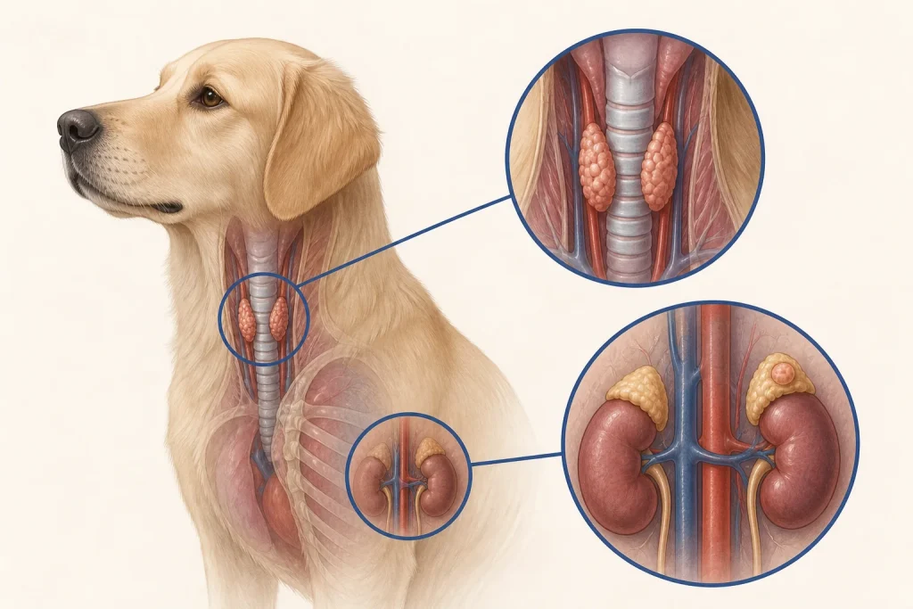
Summary
An adrenal mass can be incidental, hormone-secreting, locally invasive, metastatic, or unrelated to the dog’s current signs; endocrine testing and vascular imaging answer different questions.
Article
Evidence-informed clinical review. The discovery of an adrenal mass often creates pressure to label it benign or malignant from size alone. That shortcut is unsafe. Cortical and medullary tumors have different functional risks, and both anesthesia planning and surgical feasibility can depend on hormone activity, blood pressure, vascular invasion, metastasis, and concurrent disease.
The clinical question
The clinical team must decide whether the lesion explains the dog’s signs, whether it is functionally active, whether imaging suggests invasion into major vessels or adjacent structures, and whether the expected benefit of intervention justifies perioperative risk. An incidental finding and a crisis-producing functional tumor require different pathways.
What current evidence can establish
Endocrine tests assess hormone patterns; ultrasound and CT assess anatomy; histopathology classifies removed or sampled tissue. None is interchangeable. Tests for hyperadrenocorticism do not by themselves rule in every cortical tumor, and evaluation for pheochromocytoma requires its own clinical and biochemical reasoning. CT findings can inform, but not guarantee, resectability.
A decision-focused pathway
First confirm why abdominal imaging was obtained and whether previous studies show growth. Record blood pressure, electrolytes, kidney and liver context, medication exposure, episodic weakness, collapse, panting, agitation, or unexplained cardiovascular events. If surgery is considered, coordinate internal medicine, anesthesia, imaging, and surgical plans before admission.
A written plan should state the suspected tumor category, confidence, functional evidence, vascular findings, metastatic assessment, preparation requirements, intraoperative contingencies, and postoperative monitoring. Observation can be a reasoned choice for some lesions, but it still needs a defined imaging and clinical follow-up interval rather than indefinite neglect.
Evidence gaps and interpretation
Retrospective surgical cohorts often select dogs judged operable and therefore cannot predict risk for every adrenal mass. Imaging features overlap, small lesions may be malignant, and large lesions are not automatically functional. Evidence about one histologic type should not be transferred to another simply because both arise near the adrenal gland.
Safety, monitoring, and escalation
Collapse, severe weakness, breathing difficulty, marked abdominal pain, or acute cardiovascular instability warrants urgent care. Sampling or manipulating a functional vascular tumor may carry substantial risk. Owners should never alter steroid, blood-pressure, or endocrine medication before testing or surgery without explicit instructions from the treating team.
Keep endocrine risk and resectability on separate tracks
An adrenal lesion can matter because it produces hormones, because of its size and local behavior, or both. A quiet clinical history does not automatically exclude functional disease, while endocrine signs do not describe vessel invasion or surgical access. A structured review therefore keeps blood pressure, compatible clinical signs, relevant laboratory patterns, and appropriately selected endocrine testing distinct from the imaging assessment.
For anatomy, the useful report describes which adrenal gland is involved, the relationship to the vena cava and renal vessels, suspected thrombus or invasion, the opposite adrenal gland, regional nodes, and other abdominal lesions. Stabilization and peri-anesthetic planning may depend on the functional assessment; procedural feasibility depends on the anatomic one. When findings conflict, the uncertainty should be named and resolved in the order that most safely changes the next decision.
Questions for the veterinary team
- What makes this mass clinically relevant rather than incidental?
- Which endocrine question is each proposed test answering?
- Does CT show vascular or adjacent-organ involvement?
- What expertise and preparation are needed if surgery is chosen?
- What exact follow-up defines a safe observation plan?
Medical disclaimer: This article is educational and cannot diagnose, treat, or determine prognosis for an individual dog. It does not replace an examination or an individualized plan from a licensed veterinarian.