Canine Urothelial Carcinoma: Interpreting Urine BRAF Testing Without Skipping Anatomy
Summary
A cautious explanation of what a urine BRAF result can add in suspected canine urothelial carcinoma and why imaging and tissue context still matter.
Article
Educational medical disclaimer: This article is general education, not an interpretation of a urine test or a diagnosis. Straining without passing urine, repeated painful attempts, severe lethargy, vomiting, or abdominal distress can signal an emergency and requires immediate veterinary care.
Blood in the urine, frequent small urinations, accidents, and straining can occur with infection, stones, inflammation, prostate disease, urothelial carcinoma, or several other disorders. Because these signs overlap, a molecular result should be integrated with urinalysis, culture when indicated, imaging, anatomy, and clinical judgment. The test is evidence, not the entire case.
First answer the obstruction question
The immediate risk is whether urine can flow. Physical examination, bladder assessment, kidney values, electrolytes, and imaging may be needed urgently in a dog that cannot urinate normally. Stabilization and relief of obstruction take priority over completing a cancer workup. Owners should not wait for a send-out test if the dog repeatedly strains and produces no urine.
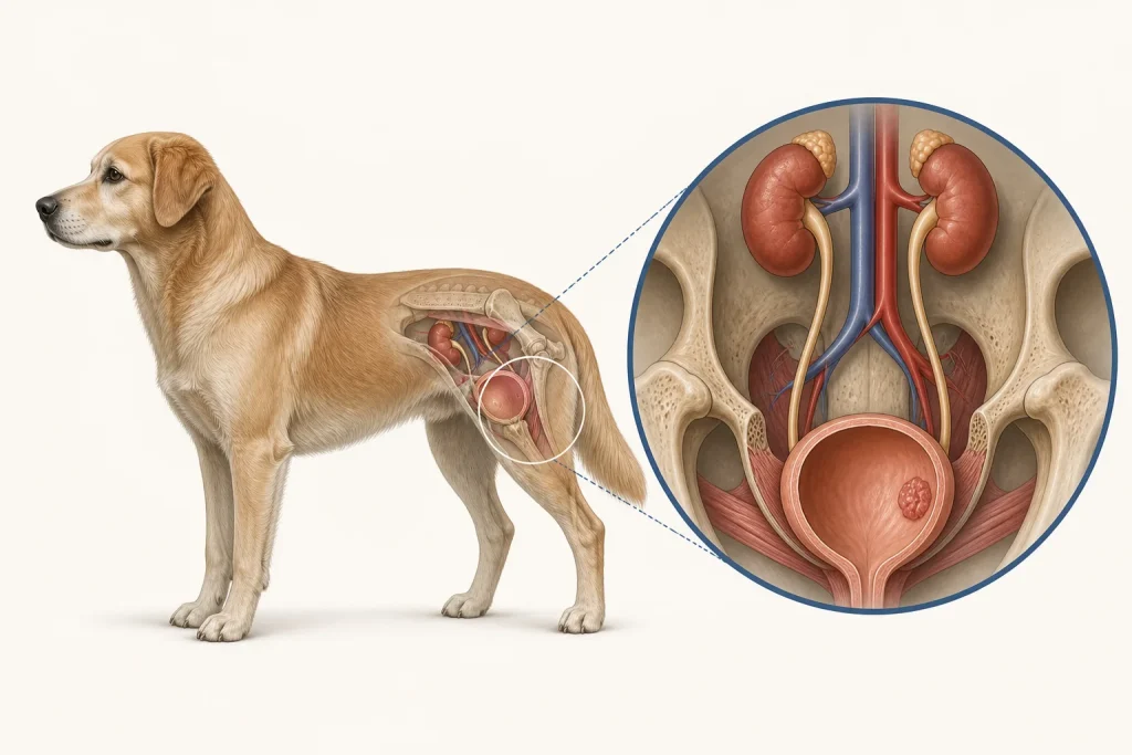
Map the urinary tract
Ultrasound or other imaging can identify a bladder or urethral lesion, show its relationship to the ureteral openings and bladder neck, evaluate kidneys and ureters, and look for stones or other structural causes. Imaging can also help select a sampling approach and establish measurements for follow-up. An apparent mass may still represent another process, while a small or difficult-to-see lesion may be clinically important because of its location.
What urine BRAF testing detects
Some canine urothelial and prostatic carcinomas carry a specific BRAF mutation that can be detected in cells shed into urine. A foundational study reported detection of the mutation in a substantial proportion of tested canine urothelial and prostatic cancers and not in the non-neoplastic comparison samples studied. A positive result in an appropriate clinical setting can therefore add meaningful support for a neoplastic process.
A negative result does not exclude carcinoma because not every tumor carries the target mutation, and specimen factors can affect detection. A later diagnostic study of urinary BRAF testing reinforces the need to interpret sensitivity and specificity in the tested population and alongside other findings. The assay also does not by itself show the exact location, size, invasiveness, grade, stage, or current obstruction risk. It should not be used to skip anatomy.
Obtaining a definitive diagnosis
Cytology or histopathology may be considered when confirmation or additional classification would change care. The collection route matters because urinary tumors can be difficult to access and clinicians consider the possibility of procedure-related tumor seeding with some techniques. Catheter sampling, traumatic catheterization, cystoscopy, surgery, or other approaches have different indications and limitations. The attending team should select the safest, highest-yield route for the lesion's location and the dog's condition.
Stage and plan by clinical purpose
Assessment may include regional lymph nodes, abdominal organs, and the thorax, selected according to diagnosis and goals. Local anatomy often drives symptoms and immediate decisions, while detectable distant disease informs prognosis and systemic-care discussions. Treatment can involve medical therapy, local interventions, radiation, surgery in selected anatomic situations, stenting or diversion for obstruction, and supportive care. No single modality is appropriate for every dog, and response should be evaluated with both symptom tracking and consistent imaging where useful.
Questions to ask after a BRAF report
- Was the result detected or not detected, and was the sample adequate?
- What does this result add to the imaging and urinalysis?
- Which alternative diagnoses remain?
- Would tissue confirmation change treatment enough to justify its risks?
- Is urine flow threatened now, and what signs require emergency care?
- How will lesion size, kidney function, infection risk, and comfort be monitored?
Owners can record urine frequency, stream quality, discomfort, visible blood, appetite, water intake, and medication timing without trying to infer tumor response from one good or bad day. Suspected infection deserves laboratory evaluation rather than automatic antibiotics, and new supplements should be disclosed because they may complicate medications or symptoms. A sound pathway treats molecular testing as one precise tool inside a broader anatomical and clinical assessment.