Canine Soft Tissue Sarcoma: Planning the Biopsy Before Planning the Margin
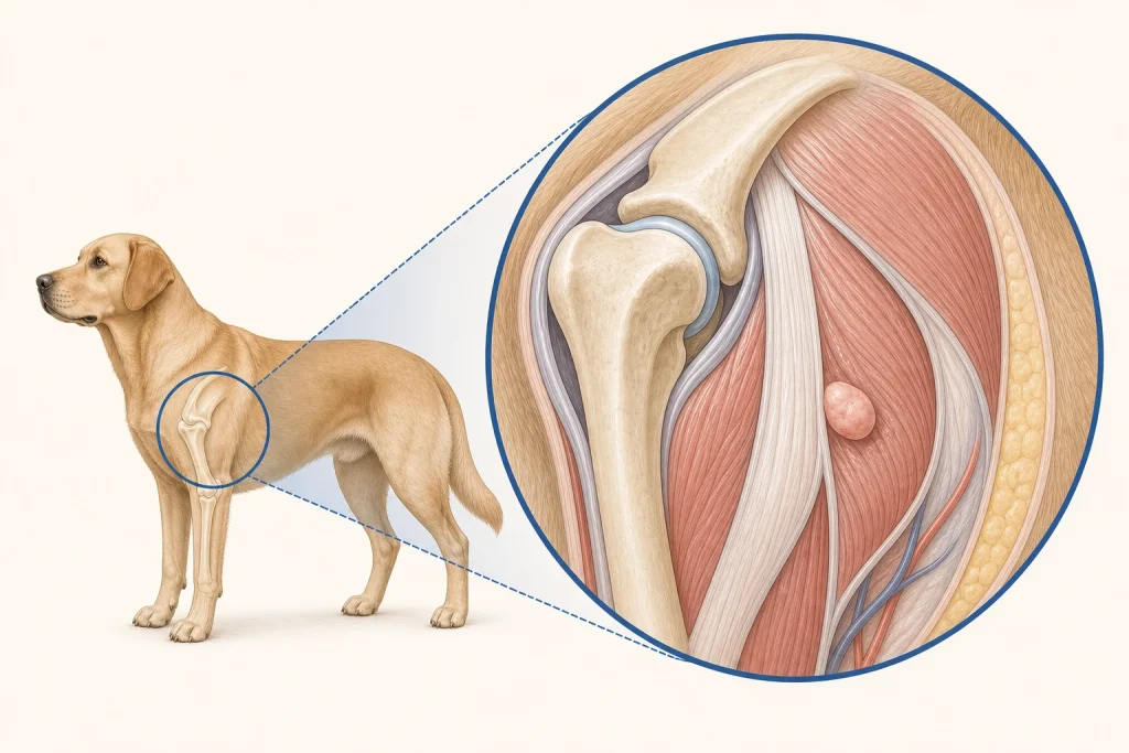
Summary
An evidence-informed pathway showing why biopsy placement, grade, anatomy, and local-control options should be coordinated before definitive surgery.
Article
Educational medical disclaimer: This review is not a diagnosis, surgical plan, or margin recommendation for any dog. A licensed veterinary team must examine the mass, review imaging and pathology, and tailor decisions to the patient.
Canine soft tissue sarcoma is a clinicopathologic grouping rather than one perfectly uniform disease. These tumors can look circumscribed from the outside while extending microscopically into surrounding planes. Their behavior varies with subtype, grade, size, location, and other factors. The most preventable planning error is treating the first operation as a simple lump removal before asking what a later definitive operation might require.
Define anatomy before the incision
The clinician should record size in three dimensions, depth, mobility, skin involvement, relation to joints or major structures, growth history, and previous procedures. A photo and body map help future comparison. Advanced imaging is not required for every superficial mass, but CT or MRI may be useful when the tumor is fixed, recurrent, large, deep, or close to critical anatomy. Imaging shows extent and guides planning; it does not replace tissue diagnosis.
A biopsy has a future footprint
A needle, core, punch, or incisional biopsy should be selected with bleeding risk, diagnostic yield, anesthesia burden, and definitive treatment in mind. The biopsy tract and scar may need to be removed with the tumor. Incisions that cross tissue compartments, create wide contamination, or compromise reconstruction can narrow later options. When a mass is large or in a complex location, early coordination with the surgeon or oncologist is valuable.
Cytology may categorize a mesenchymal process but can be poorly cellular and often cannot provide a reliable grade. Histopathology evaluates tissue architecture and may supply a specific diagnosis or the broader soft tissue sarcoma designation, plus grade when the specimen supports it. A small biopsy can occasionally underrepresent a heterogeneous tumor; the final excision specimen may refine the assessment.
Grade and margin are related but distinct
Grade summarizes selected microscopic features associated with behavior under a defined system. Margin assessment asks whether tumor reaches or approaches sampled specimen edges. Neither is absolute. Tissue shrinks after removal and fixation, specimens are sampled rather than examined molecule by molecule, and orientation affects what can be reported. A histologically complete margin reduces concern for local residual disease but does not guarantee cure. An incomplete margin raises concern but does not prove that regrowth will occur.
A systematic review and meta-analysis found higher local-recurrence risk after incomplete excision of canine cutaneous and subcutaneous soft tissue sarcomas, while also highlighting heterogeneity among studies. The 2026 veterinary oncology consensus offers a framework for diagnosis, staging, treatment, and follow-up. These sources support risk-informed planning, not a universal centimeter rule for every body site and grade.
Local-control choices
Options after diagnosis can include planned wide surgery, a more limited function-preserving operation in selected circumstances, radiation therapy, monitoring after excision, or palliative care. After an unplanned incomplete excision, scar re-excision, radiation, or surveillance may be discussed. The choice depends on grade, location, remaining anatomy, imaging, surgical morbidity, owner goals, and the evidence for that situation. Systemic therapy may be considered for selected high-risk subtypes or metastatic disease, but it is not automatically useful for every soft tissue sarcoma.
A preoperative checklist
- Has the mass been sampled, and how certain is the diagnosis?
- Would imaging change the surgical field or reconstruction plan?
- Where should the biopsy tract lie so it can be removed later?
- How will the specimen be oriented for the pathologist?
- What function or wound-healing tradeoffs accompany wider local control?
- What are the next options if margins are incomplete?
- Which follow-up findings would trigger imaging or resampling?
After treatment, owners can monitor the scar and nearby tissue with dated photographs while the veterinarian performs scheduled examinations. New swelling, wound opening, discharge, severe pain, loss of limb use, or rapid change needs earlier contact. The aim is not the widest possible intervention in every dog. It is a deliberate first plan that balances local control, function, evidence, and quality of life.